How to check bone density at home (and when to get a real test)
TL;DR: There is no FDA-cleared device that accurately measures bone density at home. What you can do is assess your risk factors, track symptoms, and run a validated tool like FRAX. A DEXA scan ordered by a clinician is the only accurate way to check bone density. It takes about 10 minutes and Medicare covers it for women over 65 and high-risk women earlier.
Can you actually check bone density at home?
The honest answer is no. Not with any accuracy that matters clinically.
There are consumer gadgets sold online that claim to estimate bone health through grip strength, ultrasound heel pads, or smartphone sensors. None of these are FDA-cleared for diagnosing osteoporosis or osteopenia, and none produce a T-score, which is the actual measurement clinicians use to diagnose bone loss. The heel ultrasound devices used in some pharmacies and health fairs are closer to real screening tools, but even they are not equivalent to a DEXA scan and should not be used to make treatment decisions [1].
What you can do at home is meaningful, though. You can calculate your personal fracture risk with validated tools, inventory your modifiable risk factors, notice physical warning signs, and decide whether to push for a formal test. That process takes about 15 minutes and beats any home gadget currently on the market.
This article walks through exactly that process, then explains what happens when you do get a real bone density test.
What is bone density and why does it drop in midlife women?
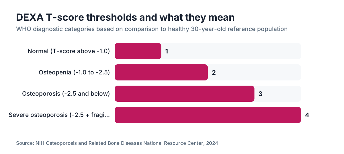
Bone density is how much mineral, mostly calcium and phosphate, is packed into a given area of bone. The clinical standard is areal bone mineral density (aBMD), measured in grams per square centimeter by dual-energy X-ray absorptiometry (DEXA). A T-score compares your density to a healthy 30-year-old reference population. A T-score at or below -2.5 is osteoporosis. Between -1.0 and -2.5 is osteopenia [2].
Estrogen is the main hormone that keeps bone remodeling in balance. Osteoclasts break old bone down; osteoblasts build new bone up. Estrogen suppresses osteoclast activity. When estrogen drops sharply, as it does during perimenopause and the first years after menopause, bone resorption outpaces formation. Women can lose 1 to 3 percent of bone mass per year in the first five years after their final period, compared to about 0.3 to 0.5 percent per year before menopause [3].
By age 65, a woman has often lost 10 to 20 percent of her peak bone mass with no obvious symptoms. That loss is silent. Osteoporosis does not hurt until a bone breaks.
What risk factors can you assess right now without a test?
This is where the real at-home work happens. Run through this list honestly.
Age and menopause timing. The earlier estrogen drops, the more cumulative bone loss. If you went through menopause before 45, your risk is higher than average [3]. If you are still in perimenopause, bone loss has likely already started.
Family history. A parent who had a hip fracture roughly doubles your own hip fracture risk, independent of your bone density [4].
Body weight. Very low body weight (BMI under 19) is a strong risk factor. Bone responds to mechanical loading; more body weight generally means denser bones. This is one reason GLP-1 medications like semaglutide have raised questions about bone health in women using them for weight loss: significant weight loss can reduce that mechanical stimulus [5].
Smoking. Current smoking lowers bone density by impairing estrogen metabolism and reducing blood supply to bone.
Alcohol. More than two drinks per day consistently suppresses osteoblast activity.
Corticosteroids. Taking prednisone or equivalent at 5 mg/day or more for three months or longer is one of the strongest secondary causes of bone loss [4].
Calcium and vitamin D intake. Most American women under-consume both. The National Institutes of Health recommends 1,000 mg of calcium per day for women 19 to 50 and 1,200 mg per day after 51. The vitamin D recommendation for adults is 600 to 800 IU per day, though many clinicians treating bone loss use higher doses based on serum 25-OH vitamin D levels [6].
Prior fracture. A fracture after age 40 from a minor fall or no trauma at all is a major red flag.
Thyroid and other conditions. Hyperthyroidism, hyperparathyroidism, celiac disease, inflammatory bowel disease, and rheumatoid arthritis all raise bone loss risk.
How do you use the FRAX tool to estimate fracture risk at home?
FRAX is a free, web-based risk calculator built by the University of Sheffield and endorsed by the World Health Organization [4]. It takes your clinical risk factors and, optionally, your femoral neck T-score, then outputs a 10-year probability of major osteoporotic fracture (hip, spine, wrist, or humerus) and hip fracture specifically.
You can access it at sheffield.ac.uk/FRAX. Select your country (use USA), enter your age, sex, height, weight, prior fracture history, parental hip fracture, smoking status, steroid use, rheumatoid arthritis diagnosis, and alcohol use. You do not need a T-score to run it. FRAX will calculate risk from clinical factors alone.
The Bone Health and Osteoporosis Foundation (BHOF, formerly the National Osteoporosis Foundation) uses FRAX thresholds to guide treatment: a 10-year hip fracture risk of 3 percent or above, or a major osteoporotic fracture risk of 20 percent or above, is where pharmacologic treatment is typically considered [4].
FRAX is not a substitute for a DEXA scan, but running it takes five minutes and hands you a defensible number to bring to your clinician. If your FRAX probability is elevated even without a T-score, that is a strong reason to push for a DEXA now rather than waiting.
One thing FRAX does not capture well is rapid perimenopausal bone loss or the effect of long-term estrogen deficiency in early menopause. In those cases, most guidelines recommend DEXA regardless of the FRAX score [3].
Are there any home devices that measure bone density?
There are three categories of consumer products marketed around bone health. None replace DEXA.
Heel quantitative ultrasound (QUS) devices. These measure how sound waves travel through the heel bone. Some pharmacies and gyms have floor-standing versions. A handful of portable consumer models exist. QUS does correlate with fracture risk in population studies, but the correlation with DEXA T-scores is weak enough that the International Society for Clinical Densitometry (ISCD) explicitly says QUS should not be used to diagnose osteoporosis or monitor treatment [1]. Think of it as a rough signal, not a diagnosis.
Grip strength dynamometers. Grip strength tracks with overall musculoskeletal health and falls risk, and low grip strength is associated with lower bone density in population data. But the correlation is far too loose to estimate a T-score from a grip reading. A dynamometer is useful for tracking sarcopenia risk over time, not bone density.
Smartphone and wearable apps. Several apps claim to estimate bone health from gait analysis, balance tests, or even microphone-based tone transmission through finger contact. None have published validation data in peer-reviewed journals that would support clinical use. Save your money.
The clearest statement on this comes from the ISCD: "Peripheral DXA, QCT, pQCT, and QUS cannot be used to monitor treatment response in patients with osteoporosis." [1] That applies to at-home devices with even lower precision.
What physical signs at home suggest you may have bone loss?
Most bone loss is truly silent. But there are signals worth noticing.
Height loss. Losing more than 1.5 inches from your peak adult height is associated with vertebral fractures, the most common osteoporotic fracture, which often happen with no acute pain event [2]. Measure yourself properly: stand barefoot against a wall, heels together, look straight ahead. Compare to your documented height from your last physical or your driver's license.
Posture change. A new forward bend or hump at the upper back (kyphosis) can point to compression fractures in the thoracic vertebrae.
Back pain without a clear injury. Sudden mid or lower back pain in a postmenopausal woman with no fall or lifting event should prompt evaluation for vertebral fracture.
A fracture from a minor event. Breaking a wrist from a fall at standing height, or a rib from a cough or a hug, in a woman over 45 is a fragility fracture until proven otherwise.
None of these confirm a diagnosis. All of them are reasons to get a formal bone density test sooner rather than later.
Who should get a DEXA scan, and when?
Guidelines differ slightly, but here is where the major organizations land.
The U.S. Preventive Services Task Force (USPSTF) recommends DEXA screening for all women 65 and older, and for postmenopausal women younger than 65 whose FRAX 10-year risk equals or exceeds that of a 65-year-old White woman with no additional risk factors (roughly 9.3 percent for major fracture) [7].
The Bone Health and Osteoporosis Foundation recommends DEXA for women at 65, postmenopausal women under 65 with risk factors, and any woman who has had a fragility fracture [4].
The North American Menopause Society (NAMS) adds that women with premature menopause (before 40) or early menopause (40 to 45) should have DEXA at the time of menopause diagnosis, not at age 65 [3].
If you are perimenopausal and your clinician is discussing hormone replacement therapy, a baseline DEXA is reasonable. Estrogen therapy is the only intervention shown to both prevent and partially restore bone density during the menopausal transition, and knowing your starting point informs the decision [3].
Medicare covers DEXA every two years for women 65 and older and for younger women who meet specific clinical criteria, including estrogen deficiency, vertebral abnormalities on X-ray, long-term steroid use, hyperparathyroidism, or monitoring after osteoporosis treatment [8].
How does hormone therapy affect bone density, and should it change your testing plan?
Estrogen is the most studied intervention for preserving bone density in perimenopausal and postmenopausal women. The Women's Health Initiative showed that women taking combined estrogen-progestin had fewer hip and vertebral fractures over the study period, with a relative risk reduction of around 33 percent for hip fracture [9].
If you are considering or already using an estrogen patch or other hormone therapy, your clinician should discuss bone density as part of that conversation. A baseline DEXA before starting lets you measure whether HRT is holding or improving your density at a follow-up scan two years later.
Progesterone may also act directly on bone through progesterone receptors on osteoblasts, though the data here are less consistent than for estrogen.
WomenRx clinicians routinely fold bone density risk assessment into hormone replacement therapy consultations, which is one practical way to formalize that conversation if your primary care provider has not brought it up.
One note on GLP-1 medications: rapid weight loss from semaglutide or tirzepatide reduces the mechanical load on bone. Some studies show modest decreases in bone mineral density with significant weight loss. If you are using semaglutide for weight loss and are already in a moderate or high FRAX risk category, discuss a DEXA with your prescriber and make sure your calcium and vitamin D intake is adequate [5].
What does a DEXA scan involve, and how do you read your results?
A DEXA scan is fast, painless, and involves very low radiation, about one-tenth the dose of a standard chest X-ray. You lie on a table while a low-dose X-ray arm passes over your hip and lumbar spine. It takes about 10 minutes. No injection, no fasting required [2].
Your results come back as two numbers for each site measured.
T-score: Compares your bone density to a young adult reference. This is the diagnostic number.
- T-score at or above -1.0: normal bone density
- T-score between -1.0 and -2.5: osteopenia (low bone mass)
- T-score at or below -2.5: osteoporosis [2]
Z-score: Compares your density to people your own age and sex. A Z-score at or below -2.0 suggests bone loss beyond what is expected for your age and warrants a search for secondary causes.
The lumbar spine and total hip (or femoral neck) are the two standard sites. If results differ between sites, management is typically based on the lower T-score.
Results alone do not dictate treatment. Your clinician combines your T-score with your FRAX probability and clinical picture to decide whether lifestyle change, supplementation, or a medication like a bisphosphonate makes sense.
| T-score range | Diagnosis | Typical next step | |---|---|---| | -1.0 and above | Normal | Rescreen in 5-10 years | | -1.0 to -2.5 | Osteopenia | Lifestyle + supplement optimization; rescreen in 2-3 years | | -2.5 and below | Osteoporosis | FRAX + consider prescription treatment | | -2.5 + fragility fracture | Severe osteoporosis | Prescription treatment strongly recommended |
What can you do at home to protect bone density starting today?
You do not need a test result to start protecting your bones. The interventions below have evidence behind them.
Weight-bearing and resistance exercise. Walking helps but is not enough on its own. Resistance training and impact activities (hiking, jogging, jumping) create the mechanical stress that stimulates bone formation. A meta-analysis in the British Journal of Sports Medicine found combined aerobic and resistance exercise significantly increased lumbar spine BMD in postmenopausal women compared to controls [10]. Aim for at least two resistance sessions per week.
Calcium from food first. Dairy, fortified plant milks, canned salmon with bones, and leafy greens like bok choy and kale are good sources. If diet alone cannot reach 1,200 mg per day after 51, a supplement fills the gap. Calcium carbonate is cheaper but needs stomach acid; take it with meals. Calcium citrate can go down any time [6].
Vitamin D. Get your 25-OH vitamin D level checked. Most labs flag deficiency below 20 ng/mL; many clinicians treating bone health aim for 30 to 50 ng/mL. The tolerable upper intake level from supplements is 4,000 IU per day for adults [6].
Stop smoking. In some analyses the bone density loss from smoking rivals early menopause.
Limit alcohol. Keep it at one drink per day or less.
Fall prevention. For women who already have osteoporosis, preventing falls matters as much as improving density. Balance training, clearing home hazards, and reviewing medications that cause dizziness all cut fracture risk.
How do you talk to your doctor about getting a bone density test?
Many primary care visits move fast, and bone density does not come up unless you raise it. Here is how to make that conversation efficient.
Before your appointment, run your FRAX score and write the number down. Note any risk factors from the list above. Measure your height at home and compare it to a prior recorded measurement.
At the appointment, say something specific: "I ran the FRAX calculator and my 10-year major fracture risk came out at X percent. I am postmenopausal and have two additional risk factors. Can we order a DEXA scan?" Specific numbers move clinical conversations faster than general concern.
If your clinician says you are too young or not high risk enough, ask which guideline they are applying. The USPSTF, BHOF, and NAMS criteria are all publicly available, and any of them may support your case depending on your age and history [3][4][7].
If you are also working with a hormone therapy provider, that is another avenue. Bone density is legitimately part of the perimenopause and menopause conversation, and a telehealth platform that handles hormones in full, as WomenRx does, can often order DEXA through your local imaging center.
How often should you recheck bone density?
The interval depends on your current results and risk factors.
For women with normal bone density and average risk, most guidelines support rescreening every 10 to 15 years [7]. A 2012 study in the New England Journal of Medicine followed women with normal baseline DEXA and found the estimated time for 10 percent of them to develop osteoporosis was 16.8 years, which suggests very infrequent rescreening is safe for women with normal density [11].
For women with osteopenia (T-score -1.0 to -2.5), the interval varies with severity. A T-score in the mild range (-1.0 to -1.5) justifies rescreening in about five years. A T-score in the -2.0 to -2.5 range warrants a two-year interval [11].
For women on osteoporosis treatment, a DEXA is typically repeated at one to two years to confirm response to therapy.
Medicare covers DEXA every two years for eligible beneficiaries, which works as a practical default for older women [8].
Frequently asked questions
Is there a home test kit for osteoporosis?
No home test kit can accurately diagnose osteoporosis. Some kits sold online claim to assess bone health through urine markers of bone turnover, but these are not validated for individual diagnosis and are not FDA-cleared for osteoporosis screening. The only accurate bone density measurement is a DEXA scan performed on medical imaging equipment, which requires a clinician referral and takes about 10 minutes at a radiology center.
Can a heel ultrasound machine at the pharmacy check my bone density?
Heel quantitative ultrasound (QUS) gives a rough signal about fracture risk and correlates modestly with bone density, but the International Society for Clinical Densitometry states QUS cannot be used to diagnose osteoporosis or monitor treatment. A result from a pharmacy QUS machine is not a T-score, cannot substitute for DEXA, and should not drive treatment decisions on its own. Use it only as a flag to seek formal DEXA testing.
What is a normal T-score for a 50-year-old woman?
A T-score at or above -1.0 is normal by WHO criteria. Many 50-year-old women fall in the osteopenia range (-1.0 to -2.5), which reflects expected bone loss from the menopausal transition rather than disease. Whether osteopenia in a 50-year-old warrants treatment depends on the FRAX fracture probability and other risk factors, not the T-score alone. A T-score of -2.5 or below at any age meets the diagnostic threshold for osteoporosis.
How accurate is the FRAX calculator without a bone density result?
FRAX without a T-score still captures most of its predictive value because most fracture risk comes from clinical factors rather than bone density alone. Studies validating FRAX show it performs reasonably well for population-level risk stratification even without densitometry input. However, FRAX tends to underestimate risk in women with rapid perimenopausal bone loss or early menopause, where a DEXA adds independent information.
At what age should a woman get her first bone density test?
The USPSTF recommends universal DEXA screening at age 65. For postmenopausal women under 65, screening is recommended if their 10-year FRAX fracture risk equals or exceeds about 9.3 percent, the level of a 65-year-old with no additional risk factors. The Bone Health and Osteoporosis Foundation and NAMS recommend earlier testing for women with premature or early menopause, fragility fractures, long-term steroid use, or other secondary causes of bone loss.
Does menopause always cause bone loss?
Menopause reliably speeds up bone loss because estrogen suppresses the osteoclasts that break down bone. When estrogen drops, resorption outpaces formation. The rate varies: some women lose 1 to 3 percent per year in the first five years after their final period; others lose less. Genetics, body weight, exercise habits, and calcium and vitamin D intake all influence how much density a given woman loses.
Can you improve bone density with exercise alone?
Exercise can slow bone loss and modestly improve density at specific sites, but it rarely reverses significant osteoporosis on its own. Weight-bearing and resistance exercise are the most effective types. A meta-analysis in the British Journal of Sports Medicine found combined aerobic and resistance training significantly increased lumbar spine BMD in postmenopausal women. Exercise matters regardless of whether medication is used, but women with osteoporosis T-scores typically need pharmacologic treatment on top of it.
Does GLP-1 weight loss affect bone density?
Significant weight loss from GLP-1 medications like semaglutide can reduce bone mineral density by decreasing the mechanical load on the skeleton. Studies of GLP-1 receptor agonists have shown modest BMD decreases at the hip with substantial weight loss. Women in perimenopause or postmenopause using GLP-1s for weight loss should keep calcium and vitamin D intake adequate and discuss baseline and follow-up DEXA with their prescriber, especially if other bone loss risk factors are present.
How much does a DEXA scan cost without insurance?
Out-of-pocket DEXA costs vary widely by geography and facility. Community imaging centers often charge $75 to $150 without insurance. Hospital-based imaging departments may charge $200 to $300 or more. Medicare covers DEXA every two years for women 65 and older and for younger women meeting specific clinical criteria. Many private insurance plans cover DEXA under the USPSTF grade B recommendation for women 65 and older and high-risk younger postmenopausal women.
What vitamins actually help bone density?
Calcium and vitamin D have the strongest evidence. The NIH recommends 1,200 mg of calcium per day for women over 51 and 600 to 800 IU of vitamin D, though clinicians often target serum 25-OH vitamin D above 30 ng/mL, which may require higher supplemental doses. Vitamin K2 comes up for bone health, but the evidence is less consistent. Adequate protein intake also supports the bone matrix.
What is the difference between osteoporosis and osteopenia?
Osteopenia and osteoporosis both describe low bone density measured by DEXA, at different thresholds. A T-score between -1.0 and -2.5 is osteopenia, meaning below-average bone mass but not yet at the fracture risk level of osteoporosis. A T-score at or below -2.5 is osteoporosis. Osteopenia does not automatically require prescription treatment; that decision depends on fracture probability from FRAX and other clinical factors.
Can hormone replacement therapy reverse bone loss?
Estrogen therapy prevents further bone loss and can partially restore density, but it is not usually described as returning established osteoporosis to normal density. The Women's Health Initiative found combined estrogen-progestin therapy reduced hip fracture risk by about 33 percent. Estrogen works best when started early in the menopausal transition. If significant osteoporosis is already present, bisphosphonates or other bone-specific agents may be needed in addition to or instead of hormone therapy.
How do I know if my back pain could be a vertebral fracture?
Vertebral compression fractures from osteoporosis often cause sudden mid or upper back pain that worsens with standing and eases with lying down. They can also happen with no pain at all. Warning signs include new back pain in a postmenopausal woman over 50 without a clear injury, height loss of more than 1.5 inches from peak, or new kyphosis. If any apply, ask your clinician for thoracic and lumbar spine X-rays in addition to a DEXA scan.
Sources
- International Society for Clinical Densitometry (ISCD), Official Positions
- NIH Osteoporosis and Related Bone Diseases National Resource Center
- The Menopause Society (formerly NAMS), position statements on bone health
- Bone Health and Osteoporosis Foundation (BHOF) and FRAX, University of Sheffield
- Endocrine Society, Pharmacological Management of Obesity Clinical Practice Guideline
- NIH Office of Dietary Supplements, Calcium Fact Sheet for Health Professionals
- U.S. Preventive Services Task Force, Osteoporosis Screening Recommendation
- CMS Medicare.gov, Bone Mass Measurement Coverage
- Women's Health Initiative, National Heart, Lung, and Blood Institute
- British Journal of Sports Medicine, exercise and bone mineral density in postmenopausal women meta-analysis
- New England Journal of Medicine, Gourlay et al. 2012, Bone-Density Testing Interval and Transition to Osteoporosis