How long does a bone density scan take (and what to expect)
TL;DR: A bone density scan (DEXA) takes about 10 to 30 minutes from the time you lie down to the time you get up. The scan itself runs 10 to 20 minutes. Check-in and positioning add a few minutes on each end. No needles, no enclosed tube, no recovery time. You can drive yourself there, get scanned, and be back at your desk by lunch.
What is a bone density scan and what does it actually measure?
A bone density scan measures how much mineral is packed into a section of bone. The test is almost always done with dual-energy X-ray absorptiometry, which everyone shortens to DEXA or DXA. The machine sends two low-dose X-ray beams through your body, usually at the hip and lumbar spine, and calculates how much energy each beam absorbs. Dense bone absorbs more. Porous bone absorbs less. The result is a number called bone mineral density (BMD), reported in grams per square centimeter. [1]
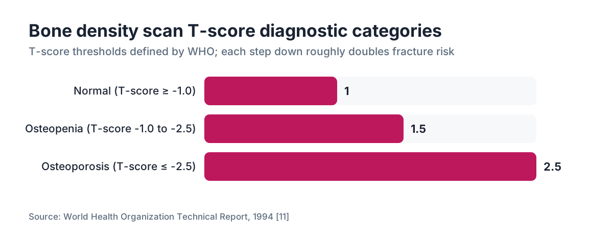
That BMD number gets compared to two reference groups. The T-score compares you to a healthy 30-year-old of the same sex. The Z-score compares you to people your own age and sex. Your doctor mostly cares about the T-score for fracture risk: at or above minus 1.0 is normal, minus 1.0 to minus 2.5 is osteopenia, and minus 2.5 or lower is osteoporosis. [2]
People mix up a DEXA with a bone scan all the time. They are different tests. A bone scan (technically bone scintigraphy) involves injecting a small amount of radioactive tracer, waiting two to four hours for it to travel through your bloodstream, then imaging your whole skeleton to look for cancer, infection, or fracture. It takes three to five hours total and uses nuclear medicine. A DEXA is purely an X-ray measurement of density, takes under 30 minutes, and involves no injections. [3] For most women asking about bone health, DEXA is the test their doctor means. See our full guide to the bone density test for more on reading your results.
How long does a bone density scan take from start to finish?
Plan for 20 to 30 minutes door to door. The scan itself runs 10 to 20 minutes, depending on whether your doctor orders imaging at just the spine, just the hip, or both. Check-in, paperwork, and the few minutes the technologist spends positioning you fill out the rest. [1]
Here is a rough breakdown of how the time is spent:
| Step | Typical time | |---|---| | Check-in and paperwork | 5 minutes | | Changing (if needed) and positioning | 3 to 5 minutes | | Spine scan | 5 to 10 minutes | | Hip scan | 5 to 10 minutes | | Technologist review before you leave | 2 to 3 minutes | | Total | 20 to 30 minutes |
If your doctor also ordered a forearm scan, add another 5 to 10 minutes. Forearm imaging is less common. Doctors order it when the hip or spine cannot be measured accurately, often because of hardware or severe arthritis. [1]
The scan is not a procedure. You lie flat on a padded table, the arm of the machine passes slowly over you, and you breathe normally. Nothing touches you except the table. There is no enclosed tube, so claustrophobia is off the table too. The radiation dose is tiny: roughly 1 to 10 microsieverts, compared to about 3 to 7 microsieverts from a chest X-ray and about 80 microsieverts from a mammogram. [4]
What age should women get a bone density scan?
Every woman 65 and older should get screened. That is the flat recommendation from the U.S. Preventive Services Task Force. For women younger than 65, the USPSTF recommends screening if their 10-year fracture risk equals or exceeds that of a 65-year-old white woman with no additional risk factors, which works out to roughly a 9.3 percent 10-year risk using the FRAX tool. [5]
Recommendations vary a little by professional society. The North American Menopause Society and the Endocrine Society both support earlier screening for postmenopausal women under 65 who carry clinical risk factors: low body weight, a fragility fracture after age 50, a family history of osteoporosis, smoking, heavy alcohol use, or long-term glucocorticoid therapy. [2][6]
Perimenopause matters here more than most women realize. Bone loss speeds up sharply in the two years before and the first few years after the final menstrual period, driven by falling estrogen. Some studies find women lose 10 to 20 percent of bone density in the decade surrounding menopause. [6] If you are in perimenopause and have risk factors, asking about a baseline DEXA in your mid-to-late 40s or early 50s is reasonable, even though it falls outside the standard screening age. To place yourself in that transition, our articles on perimenopause age and when menopause starts help.
Men are not exempt. They lose bone more slowly, and the guidelines for men are less settled. The Endocrine Society suggests considering DEXA in men 70 and older, or younger men with significant risk factors. [6]
Do you need to do anything to prepare for a bone density scan?
Prep is close to nothing. Most facilities ask you to skip calcium supplements for 24 hours beforehand, because concentrated calcium sitting in the GI tract can throw off spine measurements. Eat and drink normally otherwise. You do not fast. [1]
Wear comfortable clothing with no metal: no underwire bras, no belt buckles, no zippers over the hips or lower back. Many centers hand you a gown; some will scan you in your own clothes if they are metal-free. Leave jewelry at home so you are not fishing it out of a locker afterward.
Had a recent barium study, nuclear medicine scan, or any contrast injection? Tell the ordering physician before you schedule. Residual contrast agents can linger for days and distort the X-ray readings. General guidance is to wait at least 7 days after a barium study and several days after most other contrast agents, but your imaging center will give you a specific number. [1]
Tell the technologist if you are or might be pregnant. The dose is very low, but any X-ray during pregnancy gets minimized when it can be.
What happens during the scan and will it hurt?
It does not hurt. That is the short answer.
You lie on a flat, cushioned table. The technologist sets your feet in a triangular foam block that rotates your hips slightly inward for the hip scan, then repositions you for the spine. A mechanical arm moves slowly above you, parallel to your body. You hear a quiet hum. The arm makes one or two passes. You hold reasonably still and breathe normally. Then it is over.
Women with arthritis or back pain sometimes find the positioning mildly uncomfortable, but nothing about it is painful. The whole time you spend on the table runs 15 to 20 minutes at most. [1]
Results usually reach your ordering physician within a few days. The machine produces an image, but what the doctor reads is the numerical T-score and Z-score, plus a calculated 10-year fracture probability. Some facilities hand you a printed summary before you leave. Others post it to a patient portal.
Bone scan vs bone density scan: what is the difference?
They sound alike and answer completely different questions, which is why the mix-up is so common. A bone density scan tells you how strong your bones are. A bone scan tells you whether something is wrong inside them.
A DEXA (bone density scan) measures how dense your bones are right now. It answers one question: are you at risk for fractures? It takes 20 to 30 minutes, uses very low-dose X-ray, and needs no prep beyond skipping calcium supplements. [1]
A bone scan (bone scintigraphy, or nuclear medicine bone scan) hunts for areas of abnormal bone activity: cancer that has spread, stress fractures, bone infections, Paget's disease. You get an intravenous injection of a radioactive tracer (usually technetium-99m), wait 2 to 4 hours for it to circulate, then lie in a scanner for 30 to 60 minutes while a gamma camera images your whole skeleton. Total time commitment is 3 to 5 hours. The radiation dose runs far higher than a DEXA. [3]
If your doctor said "I'm ordering a bone density scan," they mean DEXA. If they said "bone scan" in the context of cancer staging or unexplained bone pain, they likely mean nuclear medicine. When you are unsure, ask flat out: "Is this the DEXA with the T-score, or the nuclear medicine scan with the injection?"
| Feature | DEXA (bone density) | Bone scintigraphy (bone scan) | |---|---|---| | Purpose | Measure mineral density, fracture risk | Detect cancer, infection, fracture activity | | Injection | No | Yes (radioactive tracer) | | Wait time | None | 2 to 4 hours post-injection | | Total time | 20 to 30 min | 3 to 5 hours | | Radiation dose | 1 to 10 µSv | ~4,400 µSv [3][4] | | Result format | T-score, Z-score, BMD | Whole-body image showing hot/cold spots |
How often should you repeat a bone density scan?
How often you repeat depends on your baseline result and your risk level. There is no fixed annual schedule for everyone, and the USPSTF and major societies do not push one. [5]
Women with normal bone density (T-score above minus 1.0) can generally wait 10 to 15 years if no new risk factors show up. Women with osteopenia (T-score minus 1.0 to minus 2.5) usually repeat every 1 to 5 years, with the interval tightening as the score creeps toward minus 2.5. Women already diagnosed with osteoporosis and starting medication typically repeat at 1 to 2 years to see whether treatment is working. [2][6]
Insurance coverage tracks these intervals. Medicare covers DEXA every 24 months for women who qualify, with shorter intervals allowed when medically necessary. [7] Most private insurers follow similar logic. Paying out of pocket, DEXA runs roughly $75 to $250 at most imaging centers, though hospital-based imaging can cost more.
One practical note that saves confusion later: try to repeat your scan on the same machine, or at least the same manufacturer's equipment. T-scores are not perfectly interchangeable across machines. Tracking your own trend over time requires that kind of consistency.
How does estrogen loss during menopause affect bone density?
Estrogen is the main brake on bone breakdown in women. It slows the activity of osteoclasts, the cells that dissolve old bone. When estrogen drops in perimenopause and menopause, those cells speed up and bone loss accelerates. The average woman loses 1 to 2 percent of bone mass per year in the years around her final period. Some women lose considerably more. [6]
This is why bone density age and menopause status cannot be separated. A woman who reaches menopause at 45 has roughly 5 extra years of fast bone loss compared to a woman who reaches it at 52. By the time the standard screening age of 65 arrives, that head start on loss can add up to a real gap.
Hormone replacement therapy preserves bone density. Multiple randomized trials, including the Women's Health Initiative, found that estrogen therapy cut fracture rates by roughly 33 percent at the hip and 34 percent at the spine in postmenopausal women. [8] NAMS and the Endocrine Society both recognize HRT as an appropriate option for fracture prevention in women who start it within 10 years of menopause or before age 60, when there are no contraindications. [2][6]
If you are weighing HRT for bone or other menopausal symptoms, our articles on hormone replacement therapy, the estrogen patch, and progesterone cover the options in detail. Platforms like WomenRx offer telehealth access to clinicians who focus on hormone management for women in this transition.
Can lifestyle changes actually improve your T-score?
Yes, within limits. Lifestyle changes can slow bone loss, hold onto what you have, and in some cases produce modest T-score gains. They are not a stand-in for medication once osteoporosis is established. As prevention and as a companion to treatment, they earn their keep.
The evidence is clearest for three moves. Weight-bearing exercise (walking, jogging, dancing, resistance training) stimulates bone formation; structured programs of 30 to 60 minutes most days show 1 to 3 percent improvements in spine and hip BMD. [9] Calcium of 1,000 to 1,200 mg per day for women over 50, ideally from food rather than pills, gives bone its raw material. Vitamin D of 800 to 1,000 IU daily keeps your gut absorbing that calcium. [6]
Quitting smoking matters. Smoking independently drags down BMD and raises fracture risk, and some of that risk reverses after you stop. Heavy drinking (more than 2 drinks a day, consistently) also speeds bone loss.
Falls prevention is the underrated one. Most hip fractures in older women come from falling onto a weakened hip, not from the bone simply crumbling on its own. Balance training, clearing home hazards, and reviewing medications that cause dizziness cut fracture rates even in women whose T-score never budges. [5]
Does body weight affect bone density scan results?
It does, in both directions. Higher body weight generally produces higher bone density, because bone adapts to carry more load. That is why very lean women and women with low BMI sit at higher fracture risk. It also means significant weight loss, including from GLP-1 medications, can pull bone density down if nobody is watching for it.
GLP-1 receptor agonists like semaglutide and tirzepatide are everywhere now for weight loss, and they have real metabolic benefits. But the weight they take off is not pure fat. Studies show roughly 25 to 40 percent of the weight lost on GLP-1 medications comes from lean mass, and some evidence suggests bone loss rides along with large drops in weight. [10] The clinical significance is still being worked out, and not every woman on a GLP-1 will lose meaningful bone, but it is worth raising with your prescriber, especially if you already have osteopenia or a family history of fractures.
A baseline DEXA before you start a GLP-1, and a follow-up at one to two years, is a sensible plan for women at elevated bone risk. For more on the drugs themselves, see our pieces on semaglutide and semaglutide vs tirzepatide.
How do you understand your DEXA results after the scan?
Your report lands with several numbers. Here is where to look first.
The T-score sets your clinical category. The World Health Organization drew the cutoffs: at or above minus 1.0 is normal; minus 1.0 to minus 2.5 is osteopenia; minus 2.5 or below is osteoporosis. [11] Each full standard deviation drop roughly doubles fracture risk, so a minus 2.0 carries meaningfully more danger than a minus 1.5.
The Z-score compares you to age-matched peers. A Z-score below minus 2.0 means your bone density is lower than expected for your age, which sends your doctor looking for secondary causes: thyroid disease, celiac disease, certain medications, hyperparathyroidism.
The FRAX score, often folded into the report, calculates your 10-year probability of a major osteoporotic fracture (hip, spine, wrist, or shoulder) from your T-score plus clinical risk factors. A FRAX hip fracture probability above 3 percent, or a major fracture probability above 20 percent, generally opens a treatment conversation. [5]
The BMD number itself (grams per square centimeter) earns its keep mainly for tracking change on repeat scans. A shift of more than 3 to 6 percent between scans (the exact figure depends on the machine's precision) counts as real change rather than measurement noise.
Will insurance cover your bone density scan?
Medicare Part B covers DEXA every 24 months for women who are estrogen deficient and at clinical risk for osteoporosis, who have a vertebral abnormality, who are on long-term glucocorticoid therapy, who have primary hyperparathyroidism, or who are monitoring response to osteoporosis treatment. [7]
For women under 65 with private insurance, the Affordable Care Act requires coverage without cost sharing for preventive services that earn a grade A or B from the USPSTF. DEXA for women 65 and older has a B grade, so plans must cover it for that group. For younger women, coverage hinges on whether the insurer treats the scan as preventive or diagnostic. If your doctor orders it because you have symptoms or risk factors, it may be coded as diagnostic and hit your deductible.
Out of pocket at a free-standing imaging center typically runs $75 to $150. Hospital-based imaging can hit $200 to $350 or higher for the identical scan. A few retail chains and academic medical centers have offered screening DEXAs for under $100 without insurance. Call around if cost is a concern, because prices for the same test swing wildly within one city.
WomenRx clinicians can help you sort out whether a DEXA fits your hormone history and risk profile, and can write the referral letter some insurers require for coverage.
Frequently asked questions
How long does a bone density scan take at most imaging centers?
Most women are in and out in 20 to 30 minutes total, including check-in and positioning. The actual scan runs 10 to 20 minutes, depending on whether one or two sites (spine, hip) are imaged. There is no recovery time. You can drive yourself and head straight back to work.
What should I wear to a bone density scan?
Wear loose, comfortable clothing with no metal. Skip underwire bras, jeans with metal rivets, and belts with buckles near the hip or lower back. Many centers will scan you in your own clothes if they are metal-free; others hand you a gown. Leave jewelry at home to save time.
Is a bone density scan the same as a bone scan?
No. A bone density scan (DEXA) uses low-dose X-ray to measure mineral density and takes 20 to 30 minutes with no injection. A bone scan (nuclear medicine scintigraphy) involves an IV injection of a radioactive tracer, a 2 to 4 hour wait, and 30 to 60 minutes of imaging to detect cancer, infection, or fractures. Total time for a bone scan is 3 to 5 hours.
At what age should women start getting bone density scans?
The USPSTF recommends screening for all women 65 and older. Women under 65 should be screened if their 10-year fracture risk equals or exceeds that of a 65-year-old white woman with no added risk factors (roughly 9.3 percent on the FRAX tool). Women with early menopause, low BMI, or glucocorticoid use may benefit from earlier baseline testing.
Does a bone density scan hurt?
No. You lie on a padded table and a mechanical arm passes over you. Nothing punctures, compresses, or touches you beyond the table surface. Women with hip or back arthritis sometimes find the positioning mildly uncomfortable, but the test is not painful. There is no injection, no contrast agent, and no recovery period.
What is a normal T-score on a bone density scan?
A T-score at or above minus 1.0 is normal. Minus 1.0 to minus 2.5 is osteopenia (lower than normal but not yet osteoporosis). Minus 2.5 or lower is osteoporosis. Each full point drop roughly doubles fracture risk, so the difference between minus 1.5 and minus 2.5 is more than semantics.
Can you eat and drink normally before a bone density scan?
Yes. No fasting is required. The only dietary rule is to skip calcium supplements for 24 hours before the scan, since concentrated calcium in the GI tract can interfere with spine measurements. Regular food, water, coffee, and medications are all fine. If your multivitamin contains calcium, skip that too for the 24 hours before.
How often should I repeat a bone density scan?
Women with normal results can generally wait 10 to 15 years before repeating. Women with osteopenia repeat every 1 to 5 years depending on their T-score. Women on osteoporosis treatment typically repeat at 1 to 2 years to check response. Medicare covers DEXA every 24 months for eligible women. Use the same equipment for follow-up scans to keep comparisons meaningful.
Does weight loss from GLP-1 medications like semaglutide affect bone density?
Possibly. Studies suggest 25 to 40 percent of weight lost on GLP-1 medications comes from lean mass rather than fat, and some evidence points to bone loss with large drops in weight. Women who already have osteopenia or fracture risk factors should discuss a baseline and follow-up DEXA with their prescriber before and after starting a GLP-1 medication.
Does hormone replacement therapy protect bone density?
Yes. The Women's Health Initiative found that estrogen therapy cut hip fractures by about 33 percent and vertebral fractures by about 34 percent in postmenopausal women. NAMS and the Endocrine Society recognize HRT as appropriate for fracture prevention in women who start it within 10 years of menopause or before age 60, absent contraindications.
How much does a bone density scan cost without insurance?
Out of pocket at a free-standing imaging center typically runs $75 to $150. Hospital-based imaging is often $200 to $350 or more for the same scan. Medicare Part B covers DEXA every 24 months for eligible women. If cost is a barrier, call several local imaging centers, since prices for the identical test vary widely within one city.
What is the difference between a T-score and a Z-score on a DEXA?
A T-score compares your bone density to a healthy 30-year-old of the same sex and is the number used to diagnose osteopenia or osteoporosis. A Z-score compares you to age-matched peers. A Z-score below minus 2.0 suggests your bone loss is greater than expected for your age, prompting a search for secondary causes like thyroid disease, celiac disease, or medications.
Is radiation from a DEXA scan dangerous?
No. The dose from a DEXA is roughly 1 to 10 microsieverts, comparable to a few hours of normal background radiation. A chest X-ray delivers about 3 to 7 microsieverts and a mammogram about 80 microsieverts. The dose is considered negligible for any woman who is not pregnant.
Can I get a bone density scan during perimenopause?
Yes, and it may be worth doing if you have risk factors. Standard guidelines recommend screening at 65, but a baseline scan in your late 40s or early 50s during perimenopause makes sense if you have low BMI, a fragility fracture history, a strong family history of osteoporosis, or take bone-depleting medications. Discuss timing with your doctor based on your profile.
Sources
- RadiologyInfo.org (ACR/RSNA) — Bone Densitometry (DEXA, DXA)
- North American Menopause Society (NAMS) — 2021 Position Statement on Osteoporosis
- Society of Nuclear Medicine and Molecular Imaging (SNMMI) — Bone Scan
- U.S. Food and Drug Administration — Medical X-ray Imaging and Radiation Dose
- U.S. Preventive Services Task Force — Osteoporosis to Prevent Fractures: Screening (2018)
- Endocrine Society — Clinical Practice Guideline: Osteoporosis in Postmenopausal Women (2019)
- Centers for Medicare and Medicaid Services (CMS) — Bone Mass Measurement Coverage
- Women's Health Initiative (WHI) — JAMA 2002; 288(3):321-333
- National Institutes of Health — Office of Dietary Supplements: Calcium Fact Sheet for Health Professionals
- New England Journal of Medicine — STEP 1 Trial: Semaglutide and Body Composition (Wilding et al., 2021)
- World Health Organization — Assessment of Fracture Risk and Its Application to Screening for Postmenopausal Osteoporosis (1994 Technical Report)