Estrogen receptor positive fat cells and menopause weight gain
TL;DR: Fat cells express estrogen receptors (mainly ERα and ERβ). When estrogen falls during menopause, those receptors lose their ligand, triggering less fat breakdown, more visceral fat storage, and lower resting metabolic rate. Studies show menopausal women gain roughly 1.5 kg of fat per year in the first two years after their final period, with the shift from subcutaneous to visceral fat being the most metabolically dangerous change.
What are estrogen receptor positive fat cells and why do they matter?
Fat cells, technically called adipocytes, are not passive storage bags. They are metabolically active tissue that expresses estrogen receptors the same way breast cells do. The two main receptors are estrogen receptor alpha (ERα) and estrogen receptor beta (ERβ), with ERα being the dominant driver of fat distribution in women [1].
When estrogen binds to ERα in adipocytes, it signals the cell to favor lipolysis (fat breakdown) over lipogenesis (fat storage), particularly in visceral depots around the abdominal organs. It also regulates adiponectin, a hormone released by fat cells that improves insulin sensitivity and keeps systemic inflammation lower. So when estrogen is present and binding well, fat cells behave differently than when estrogen is gone.
The phrase "estrogen receptor positive fat cells" is not a clinical diagnosis the way ER-positive breast cancer is. It simply describes the normal biology of adipose tissue in women: your fat cells have estrogen receptors, they respond to estrogen, and their behavior changes materially when estrogen supply drops.
This matters because menopause is the largest estrogen withdrawal most women will ever experience. The fat cells do not disappear. They just stop getting the hormonal signal that kept them working in a particular way.
How does falling estrogen at menopause change what fat cells do?
Estrogen controls fat cell function through at least three pathways that are well documented in human studies [2].
First, ERα suppresses lipoprotein lipase (LPL) activity in visceral adipose tissue. LPL is the enzyme that pulls fat from the bloodstream into a fat cell for storage. When estrogen drops, LPL activity in visceral fat rises, meaning more dietary fat and circulating triglycerides get deposited around the organs rather than in subcutaneous (under the skin) depots.
Second, estrogen influences the number and sensitivity of beta-adrenergic receptors on fat cells. Those receptors are what adrenaline and noradrenaline use to trigger fat release. Lower estrogen means fewer functional beta-adrenergic receptors in abdominal fat, so the same amount of exercise or caloric deficit produces less fat mobilization from the belly.
Third, fat cells themselves are a minor source of estrogen after menopause. An enzyme called aromatase inside adipocytes converts adrenal androgens into estradiol. This is why adipose tissue volume matters post-menopause: more fat tissue means slightly more peripheral estrogen conversion. That sounds protective, but higher adiposity also means more systemic inflammation and higher estrogen levels that carry their own risks for hormone-sensitive cancers [3].
The net result of all three mechanisms: fat accumulates preferentially in the abdomen, less gets burned at rest, and the metabolic profile of the fat cell itself becomes more inflammatory.
How much weight do women actually gain during menopause, and how much is from fat cell changes?
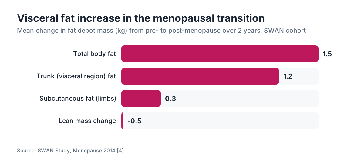
Here is where the data gets more careful. Average weight gain during midlife in women runs roughly 0.5 to 1.5 kg per year, but the Study of Women's Health Across the Nation (SWAN) found the menopausal transition itself added about 1.5 kg of body fat in the first two years after the final menstrual period, separate from aging and lifestyle [4].
Waist circumference grew even in women whose total weight held steady. That is the estrogen receptor story showing up in real bodies: the fat is not necessarily more; it is somewhere different and metabolically worse.
| Fat depot | Pre-menopause behavior | Post-menopause behavior | |---|---|---| | Subcutaneous (hips, thighs) | Preferred storage site | Less active; some redistribution | | Visceral (abdomen, organs) | Actively suppressed by ERα | Expands; LPL activity rises | | Intramuscular fat | Modest | Increases with muscle loss | | Liver fat | Low in most women | Rises, raising NAFLD risk |
The visceral shift is not cosmetic. Visceral fat secretes inflammatory cytokines including IL-6 and TNF-α, raises fasting insulin, and is independently linked to cardiovascular disease risk, which climbs sharply after menopause [5].
So the honest answer: menopausal women gain fat because of aging, reduced activity, and caloric surplus, but the estrogen receptor mechanism is why the fat lands in the worst possible place even when total weight gain is modest.
Do fat cells change how they express estrogen receptors as women age?
Yes, and this is underappreciated. ERα expression in visceral adipose tissue declines with age in women, separate from the decline in circulating estrogen [6]. That means even if you replaced estrogen, the receptor landscape in older fat cells is not identical to what it was at 35.
Some research suggests that obesity itself downregulates ERα in adipose tissue, creating a feedback loop where excess fat leads to less ERα, which leads to worse fat regulation, which perpetuates adiposity. A 2018 study in Molecular Metabolism found that ERα expression in human subcutaneous adipocytes inversely correlated with BMI, suggesting that higher fat mass blunts the very receptor that would otherwise help manage it [6].
ERβ tells a different story. It appears to have anti-proliferative effects in fat tissue, may reduce adipocyte size, and its role is still being worked out. Most of the weight gain and redistribution biology centers on ERα.
The practical implication: the longer the window of low estrogen exposure post-menopause, the more the receptor-level changes compound the hormonal ones. This is one reason clinicians who follow the menopause society guidelines often discuss the timing hypothesis, the idea that starting menopausal hormone therapy earlier in the transition may preserve metabolic benefits better than starting years later.
Does menopausal hormone therapy (MHT) reverse the fat cell changes?
Partially, and the data is more encouraging than the post-2002 WHI panic suggested. The Women's Health Initiative randomized trial, when re-analyzed by age group, showed that women who started combined estrogen-progestogen therapy within 10 years of menopause had less visceral fat accumulation than those on placebo, though total weight was similar between groups [5].
Observational and smaller randomized studies have consistently shown that oral and transdermal estradiol reduce visceral fat accumulation, improve fasting insulin, and lower LPL activity in abdominal adipose tissue. Transdermal delivery specifically avoids the first-pass liver effect that oral estrogen produces, which matters because oral estrogens raise triglycerides and sex hormone binding globulin in ways transdermal formulations largely do not [7].
Progestogen type matters too. Synthetic progestins (like medroxyprogesterone acetate) tend to blunt some of estrogen's favorable metabolic effects on fat cells, while micronized progesterone (Prometrium) appears more metabolically neutral. This is not settled science, but the clinical trend toward using body-identical micronized progesterone alongside estradiol reflects these concerns.
What MHT does not do: it does not produce weight loss. Women starting MHT should not expect the scale to drop. What it can do is slow visceral fat accumulation and hold the metabolic profile of fat cells closer to premenopausal function, which is a different and clinically meaningful benefit.
If you are exploring hormone therapy options, telehealth platforms like WomenRx can evaluate your full hormonal picture and discuss whether MHT is appropriate for your situation.
How do GLP-1 receptor agonists interact with estrogen receptor positive fat cells?
GLP-1 receptor agonists like semaglutide (Ozempic, Wegovy) and tirzepatide (Mounjaro, Zepbound) are now being used in large numbers of perimenopausal and postmenopausal women, and the question of how they interact with estrogen-deficient fat cells is genuinely interesting.
GLP-1 receptors are expressed in adipose tissue, and GLP-1 agonists appear to preferentially reduce visceral fat over subcutaneous fat, which maps almost perfectly onto the problem that estrogen receptor loss creates [8]. The SURMOUNT-1 trial of tirzepatide showed that participants lost an average of 20.9% of body weight at the highest dose, with significant reductions in waist circumference [9]. While SURMOUNT-1 was not stratified by menopausal status, post-hoc analyses suggest women over 50 had similar or slightly greater percentage fat mass reductions.
Semaglutide, as covered in semaglutide news, also reduces visceral adiposity independent of total weight loss, which makes it a particularly interesting tool for postmenopausal women whose central fat accumulation is the primary metabolic threat. For more on whether the brand-name formulations differ, the is semaglutide the same as ozempic explainer covers that directly.
The open question is whether combining MHT with a GLP-1 agonist produces additive benefits on visceral fat. No large randomized trial has tested this combination directly. The closest data comes from mechanistic studies showing that estrogen and GLP-1 signaling converge on some of the same hypothalamic circuits that regulate energy balance, and that estrogen may enhance GLP-1 receptor sensitivity. That is promising but not yet proof.
Does losing weight during menopause change estrogen receptor expression in fat cells?
This is one of the more hopeful findings in recent adipose biology. Weight loss, even without hormone therapy, appears to raise ERα expression in subcutaneous adipose tissue. A 2020 study in the Journal of Clinical Endocrinology and Metabolism found that caloric restriction over 12 weeks increased ERα mRNA expression in subcutaneous fat biopsies from postmenopausal women, suggesting the receptor capacity is not permanently lost, it is suppressed by the metabolic environment of excess adiposity [10].
This creates a positive feedback opportunity. Losing visceral fat reduces systemic inflammation, which improves receptor sensitivity. Improved receptor sensitivity means the fat cells respond better to whatever estrogen is circulating (either from the adrenal-to-aromatase pathway or from MHT). Better receptor function then helps maintain healthier fat distribution going forward.
The catch is that losing fat during menopause is harder than at younger ages for reasons that are partially the estrogen receptor story itself: lower beta-adrenergic sensitivity, higher LPL activity, and a resting metabolic rate that drops roughly 50-100 kcal per day at menopause onset beyond what aging alone predicts. That metabolic drop is real and it means the caloric math that worked at 38 does not automatically work at 52.
Exercise type matters here. Resistance training preserves muscle, which supports resting metabolic rate and improves insulin sensitivity in fat cells. High-intensity interval training appears to preferentially reduce visceral fat in postmenopausal women compared to moderate continuous exercise. Neither replaces the hormonal changes, but both improve the cellular environment in which estrogen receptors operate.
What is the connection between estrogen receptor positive fat cells and breast cancer risk?
This is where the fat cell biology intersects with a legitimate health concern that women and clinicians both carry, especially when considering hormone therapy.
Postmenopausal adipose tissue produces estrogen through aromatase. The more fat tissue, the more peripheral estrogen production. Because breast cancer is often ER-positive (meaning it is fueled by estrogen), higher adiposity after menopause raises circulating estradiol levels that can stimulate ER-positive tumor cells [3]. The American Cancer Society and the National Cancer Institute both note that obesity after menopause is an established risk factor for postmenopausal breast cancer, with relative risk estimates in the 1.2 to 1.5 range depending on degree of adiposity [3].
This creates a tension: fat cells with functional estrogen receptors help regulate healthy fat distribution, but excess adipose tissue generating estrogen outside of normal ovarian control is a risk factor for ER-positive breast cancer.
MHT adds exogenous estrogen into this system. Combined estrogen-progestogen therapy for more than five years is associated with a small but real increase in breast cancer risk. The absolute numbers matter: the WHI found roughly 8 additional breast cancers per 10,000 women per year on combined therapy compared to placebo. Estrogen-only therapy (for women who have had a hysterectomy) showed no significant increase and in some analyses showed lower breast cancer incidence.
None of this means MHT is wrong for most women. It means the conversation requires weighing these numbers against the cardiovascular and metabolic harms of untreated visceral fat accumulation, which also carry real morbidity.
How does perimenopause affect fat cells differently from full menopause?
Perimenopause gets less attention in this conversation but the fat cell changes begin well before the final menstrual period. In the years before menopause, estradiol levels fluctuate wildly rather than simply declining. This estrogen variability, more than low estrogen, disrupts the signaling that fat cells depend on [11].
During a perimenopausal low-estrogen phase, ERα-mediated suppression of visceral LPL activity relaxes, fat moves toward the abdomen, inflammation ticks up. Then estrogen surges again and some of that is reversed. Over several years of this cycling, the net trend is toward visceral accumulation and worse metabolic markers, even in women who are still having periods.
Many women notice weight gain or belly fat changes in their mid-to-late 40s while they are still peri menopausal, long before they would be classified as postmenopausal. This is the fat cell story playing out in perimenopause, more than after it.
The challenge for clinicians is that standard hormone panels during perimenopause are notoriously hard to interpret because FSH and estradiol fluctuate dramatically day to day. A single measurement showing normal estradiol does not rule out the periods of estrogen deficiency that are driving fat cell changes in the background.
What can you actually do about estrogen-driven fat changes during menopause?
There is no single answer here, and anyone who tells you otherwise is oversimplifying. The practical toolkit has a few well-supported tools and several that are popular but weakly evidenced.
Menopausal hormone therapy is the most direct intervention for preserving estrogen receptor signaling in fat cells. For women without contraindications, current guidance from the Menopause Society (formerly NAMS) and the Endocrine Society supports MHT for symptomatic menopausal women under 60 or within 10 years of menopause [12]. It slows visceral fat accumulation but should not be expected to produce weight loss.
GLP-1 receptor agonists preferentially reduce visceral fat and are increasingly prescribed for perimenopausal and postmenopausal women with obesity or weight-related metabolic risk. The data on visceral fat reduction is strong. The long-term data specifically in postmenopausal women is still accumulating.
Resistance training two to three times per week is the most accessible intervention. It preserves lean mass, maintains resting metabolic rate, improves insulin sensitivity in adipose tissue, and reduces visceral fat on its own. A 2022 meta-analysis in Obesity Reviews found resistance training reduced visceral adipose tissue by an average of 6.1% in postmenopausal women across 28 trials.
Sleep quality matters more than most weight conversations admit. Short sleep duration raises cortisol and ghrelin, both of which drive visceral fat accumulation. Menopause-related sleep disruption (hot flashes, night sweats, anxiety) is therefore more than a comfort issue; it actively worsens the fat cell environment.
Dietary protein adequacy helps preserve muscle during the period of fat redistribution. Roughly 1.2 to 1.6 grams of protein per kilogram of body weight is a range with reasonable support in women over 50, though individual needs vary.
WomenRx offers clinician-guided hormone and GLP-1 programs built for women working through these changes, if you want a starting point for a personalized conversation.
What I would not spend money on: most supplements marketed specifically for menopausal metabolism have thin or no randomized trial evidence. Phytoestrogens like soy isoflavones have very weak ERα agonist activity and have not shown meaningful visceral fat reduction in well-controlled trials.
Are there other hormones that affect fat cell receptors during menopause?
Estrogen gets the most attention, but several other hormonal changes at menopause also affect adipocyte biology, and they interact.
Insulin sensitivity drops at menopause, partly because of estrogen loss (ERα in fat cells supports insulin signaling) and partly because of the visceral fat expansion itself. More visceral fat means more free fatty acid spillover into the portal circulation, which drives hepatic insulin resistance. This creates the metabolic syndrome pattern many women develop in their 50s even if they never had insulin issues before.
Testosterone in women also affects fat cell receptors. Androgen receptors in fat cells respond to testosterone, which naturally rises relative to estrogen after menopause (because estrogen falls more than testosterone does). Higher androgen relative to estrogen favors android (abdominal) fat distribution, which compounds the ERα-loss effect [13].
Thyroid hormone status is worth checking in any woman gaining unexplained weight at midlife. Thyroid function affects metabolic rate and fat cell activity independently of estrogen. You can read more about thyroid hormone replacement therapy if that intersection applies to your situation.
Cortisol deserves a mention. Chronic stress raises cortisol, which directly promotes visceral fat storage via glucocorticoid receptors on adipocytes. Menopause-related anxiety and sleep disruption often raise average cortisol exposure. So the stress-fat-hormone feedback loop is a real contributor, though separating it from the estrogen receptor mechanism specifically is difficult in human studies.
Frequently asked questions
Do all fat cells in women have estrogen receptors?
Most human adipocytes express at least some ERα or ERβ, but receptor density varies by fat depot. Visceral adipose tissue (belly fat) and subcutaneous fat express ERα at different levels, with visceral fat being more sensitive to estrogen's suppressive effects on LPL activity. Breast adipose tissue also expresses both receptor types, which is relevant for breast cancer risk discussions.
Can you test your fat cells' estrogen receptor status at home or at a regular clinic?
No. Estrogen receptor expression in adipose tissue is measured through biopsy and laboratory analysis, which is a research procedure, not a routine clinical test. What you can test clinically are circulating estradiol levels, FSH, and metabolic markers like fasting insulin and visceral fat via DEXA scan. Those give you an indirect picture of how your fat cells are functioning.
Is belly fat after menopause purely hormonal or does aging matter too?
Both. Aging independently reduces muscle mass and slows fat oxidation regardless of hormone status. The SWAN study showed menopause itself adds visceral fat accumulation beyond what aging alone explains, but the two effects compound each other. Separating them is nearly impossible in living women, which is why clinical studies try to control for age when measuring menopause-specific changes.
Does testosterone therapy help with fat distribution after menopause?
The data is mixed. Testosterone added to estrogen therapy in postmenopausal women shows some benefit for lean mass preservation and libido, but evidence for meaningful visceral fat reduction specifically is limited. Very high testosterone can worsen android fat distribution. The Endocrine Society does not currently recommend testosterone for weight management in women, though off-label use is common.
Why do some women gain no weight during menopause while others gain a lot?
Genetics account for a substantial portion of the variance. ERα gene polymorphisms, differences in aromatase activity, baseline insulin sensitivity, sleep quality, stress load, activity level, and dietary patterns all interact. Women who maintain high physical activity and adequate protein intake through perimenopause tend to show smaller visceral fat gains, though even active women often see some redistribution.
Does the weight gain from menopause ever stop on its own?
The rapid visceral fat accumulation that occurs in the first two to three years after the final menstrual period does tend to slow as the body adapts to its new hormonal environment. However, the metabolic changes in fat cells are not fully reversed by time alone. Without intervention, the lower resting metabolic rate and changed fat distribution tend to persist.
Can bioidentical hormones restore normal fat cell function better than synthetic hormones?
The term 'bioidentical' refers to chemical structure matching the body's own hormones, which includes FDA-approved transdermal estradiol patches and micronized progesterone capsules. These do appear to have more favorable metabolic profiles than older synthetic versions. Compounded bioidenticals from pharmacies are not FDA-approved and have variable dosing accuracy; they are not demonstrably better than regulated formulations.
Does menopause-related fat gain affect cancer risk beyond breast cancer?
Yes. Postmenopausal visceral fat accumulation is associated with higher risk for endometrial cancer (through excess peripheral estrogen from aromatase in fat cells), colorectal cancer, and possibly ovarian cancer. The mechanisms involve both elevated estrogen levels and the pro-inflammatory cytokines that visceral adipose tissue secretes, particularly IL-6 and TNF-alpha.
What does a DEXA scan show about menopausal fat redistribution?
DEXA (dual-energy X-ray absorptiometry) can distinguish visceral adipose tissue from subcutaneous fat and measure regional fat mass in the trunk versus limbs. It also measures lean mass, which helps identify sarcopenic obesity (high fat plus low muscle, common in postmenopausal women). It is more informative than BMI alone and is available at many endocrinology and radiology centers.
Is the fat gained during menopause harder to lose than fat gained at other life stages?
Evidence suggests yes, though not because fat cells become irreversibly different. The lower resting metabolic rate, higher LPL activity in visceral depots, and reduced beta-adrenergic receptor sensitivity mean the same caloric deficit mobilizes less visceral fat than it would have at younger ages. GLP-1 agonists and structured resistance training are the best-evidenced tools for overcoming this resistance.
Do hot flashes have anything to do with fat cell estrogen receptors?
Hot flashes are driven primarily by hypothalamic thermoregulation changes caused by estrogen withdrawal, not directly by fat cell receptor status. However, visceral fat mass correlates with hot flash severity in observational studies, possibly because adipose tissue generates heat and inflammatory signals that lower the thermoregulatory threshold. Treating hot flashes with MHT also secondarily improves the fat cell hormonal environment.
Is there any evidence that phytoestrogens from food or supplements activate fat cell estrogen receptors meaningfully?
Phytoestrogens like genistein and daidzein (from soy) bind ERβ more than ERα and with much lower affinity than estradiol. Clinical trials of soy isoflavone supplements have not shown meaningful reductions in visceral fat or weight in postmenopausal women. Dietary soy is safe and nutritious but should not be expected to replicate estrogen's effects on fat cell biology.
How is the fat cell change in menopause different from what happens in men at midlife?
Men also accumulate visceral fat with age, driven partly by declining testosterone rather than estrogen. Men do have small amounts of estrogen (produced via aromatase in fat cells and testes) and ERα in adipose tissue, but they lack the dramatic estrogen withdrawal that menopausal women experience. The speed and magnitude of visceral fat shift at menopause is steeper than in age-matched men over the same decade. More on do men go through menopause.
Sources
- Endocrine Reviews, Mayes & Watson 2004 – Estrogen Receptor Alpha and Adipose Biology
- Journal of Clinical Endocrinology & Metabolism – Estrogen regulation of LPL and adipose metabolism
- National Cancer Institute – Obesity and Cancer fact sheet
- Study of Women's Health Across the Nation (SWAN) – Sternfeld et al., Menopause 2014
- Women's Health Initiative – Manson et al., NEJM 2003 and subsequent re-analyses
- Molecular Metabolism, Ström et al. 2018 – ERα expression and BMI in human adipocytes
- Menopause Society (NAMS) – Position statement on hormone therapy 2022
- Diabetes, Obesity and Metabolism – GLP-1 receptor expression in adipose tissue review
- New England Journal of Medicine – Jastreboff et al., SURMOUNT-1 trial, 2022
- Journal of Clinical Endocrinology & Metabolism – ERα expression after weight loss in postmenopausal women, 2020
- Climacteric – Perimenopause fat redistribution and estrogen fluctuation review
- Endocrine Society – Clinical Practice Guideline: Menopausal Hormone Therapy 2015
- Journal of Steroid Biochemistry and Molecular Biology – Androgens, androgen receptors, and adipose distribution in women