Bone mineral density test: what it measures, who needs it, and what your results mean
TL;DR: A bone mineral density (BMD) test, usually a DEXA scan, measures how much calcium and mineral is packed into your bones. It takes 10 to 30 minutes, uses very low radiation, and gives you a T-score. A T-score at or below -2.5 means osteoporosis. Women should get their first test at age 65, or earlier if risk factors like early menopause or long-term steroid use apply.
What is a bone mineral density test and what does it actually measure?
A bone mineral density test measures how much mineral, mostly calcium, is packed into a set volume of bone. More mineral means denser, stronger bone. The result comes out in grams per square centimeter (g/cm²), and that raw number gets compared against a reference population to produce two scores: the T-score and the Z-score.
The T-score compares your bone density to the average peak bone mass of a healthy 30-year-old of the same sex. The Z-score compares you to people your own age and sex. Both have their uses, but for diagnosing osteoporosis or osteopenia in postmenopausal women, the T-score is the number clinicians reach for.
Bone is living tissue. Cells called osteoclasts break it down, cells called osteoblasts rebuild it, and the two run in balance for most of adult life. Estrogen keeps osteoclasts in check. When estrogen falls during perimenopause and menopause, osteoclasts start winning, and density drops. A BMD test catches that shift before a bone breaks.
The test doesn't measure bone quality, flexibility, or microarchitecture. Two women can post the same T-score and still carry different fracture risk depending on bone geometry and turnover rate. That's why your clinician reads the number alongside your clinical risk factors instead of treating it in isolation.
How do they do a bone density test? What happens during the scan?
The standard method is dual-energy X-ray absorptiometry, almost always called a DEXA scan (sometimes spelled DXA). You lie fully clothed on a padded table. A mechanical arm passes slowly over your body, sending two low-energy X-ray beams through the bones at your hip and lumbar spine. The machine measures how much of each beam the bone absorbs, calculates the mineral content, and generates your BMD score. Start to finish takes 10 to 30 minutes depending on the equipment and how many sites get scanned [1].
Radiation exposure is tiny, around 1 to 10 microsieverts per scan. A standard chest X-ray delivers about 100 microsieverts. You absorb roughly 10 microsieverts just flying across the country. The DEXA dose sits well below any threshold of concern [2].
You don't need to fast. Skip calcium supplements for 24 hours beforehand, because a high dose sitting in the GI tract can nudge the reading. Tell the technician if you've had a recent contrast dye study or nuclear medicine scan. Metal hardware from hip replacements or spinal fusions in the scan field throws off accuracy, so the technician may switch which sites they measure.
Peripheral DEXA devices scan the wrist, finger, or heel and show up sometimes at pharmacies or health fairs. They're cheaper and faster, and they're not diagnostic. A heel result flagging low density should always get confirmed with a central DEXA of the hip and spine before anyone makes a treatment decision [3].
Quantitative CT (QCT) is another option. It gives a true volumetric density measurement and can separate trabecular bone (the spongy inner layer, which responds faster to hormonal changes) from cortical bone. QCT costs more and delivers higher radiation than DEXA, so it stays reserved for specific research or clinical situations rather than routine screening.
What do your T-score and Z-score results mean?
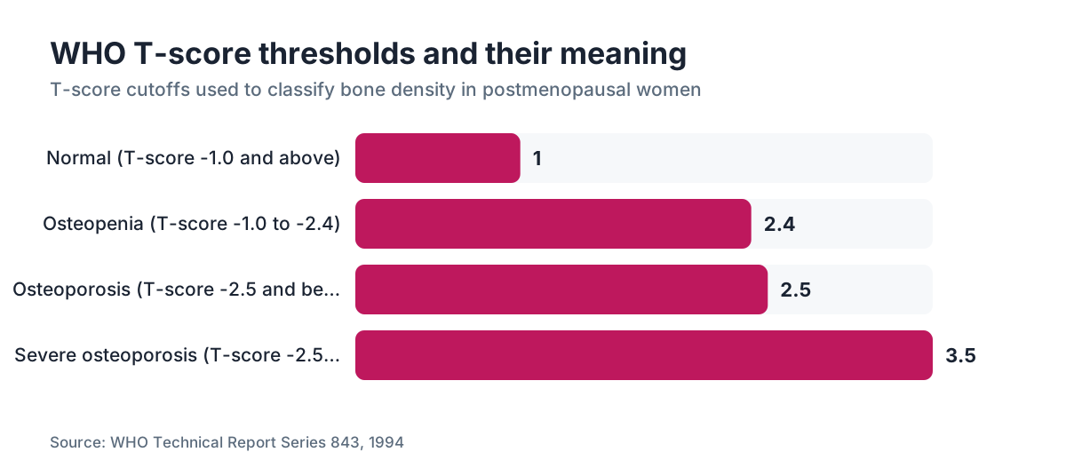
The World Health Organization set the T-score classification in 1994, and it's still the clinical standard [4].
| T-score range | Classification | |---|---| | -1.0 and above | Normal bone density | | -1.0 to -2.4 | Osteopenia (low bone mass) | | -2.5 and below | Osteoporosis | | -2.5 and below with fragility fracture | Severe osteoporosis |
Each unit drop in T-score works out to roughly 10 to 12 percent lower bone density than the reference peak. Here's the number that matters more: each standard deviation decrease in hip BMD is tied to roughly a doubling of hip fracture risk, per the analysis cited in the Bone Health and Osteoporosis Foundation clinician's guide [5].
Osteopenia is not a disease. It's a zone where bone loss has started but hasn't crossed the fracture threshold. Plenty of women in their 50s land here after menopause. Whether to treat osteopenia depends on the full FRAX score (a 10-year fracture probability calculator from the University of Sheffield), not the T-score by itself.
The Z-score tells a different story. A Z-score below -2.0 means your bone density is unusually low for your age, a signal that something beyond normal aging is driving the loss. Worth investigating: undiagnosed celiac disease, hyperparathyroidism, long-term glucocorticoid use, premature ovarian insufficiency. If your Z-score is low, your doctor should hunt for a secondary cause before writing a treatment plan.
One more wrinkle: T-scores are site-specific. Your hip and spine don't always agree. The lower of the two scores drives the diagnosis. Some women show a normal spine score and an osteoporotic hip score, partly because vertebral bone can look artificially dense on DEXA when arthritis or calcification is present. A trained radiologist reading the raw images catches that.
Who should get a bone mineral density test and at what age?
The U.S. Preventive Services Task Force (USPSTF) recommends BMD screening for every woman age 65 and older. For women under 65, screening is recommended if their 10-year fracture risk equals or exceeds that of a 65-year-old white woman with no additional risk factors, which lands around a 9.3 percent 10-year major osteoporotic fracture risk on FRAX [6].
The Bone Health and Osteoporosis Foundation and the North American Menopause Society (NAMS) add clinical judgment and recommend earlier testing if any of these apply [7]:
- Menopause before age 45 (early menopause or premature ovarian insufficiency)
- More than three months of glucocorticoid use (prednisone 5 mg/day or equivalent)
- A parent with a hip fracture
- Current smoking
- Body weight under 127 pounds (58 kg)
- Heavy alcohol use (more than 3 drinks per day)
- Rheumatoid arthritis
- Conditions causing malabsorption, including Crohn's disease and celiac disease
- History of eating disorders with significant weight loss
Men don't get tested routinely until age 70, unless risk factors are present. Testing children or teenagers is reserved for specific medical conditions and is never routine.
If you're in perimenopause and wondering whether it's too early, here's the honest answer. A baseline scan in your late 40s or early 50s gives you a personal reference point. Test again in two to five years and you can see your own rate of change, which tells you far more than a single number ever will. Not every insurer covers an early scan without a documented indication, so check your coverage before you book.
How does menopause affect bone density, and how fast do women lose bone?
Bone loss in women isn't a slow, steady slide. It speeds up sharply during the menopause transition and stays elevated for several years after. The best data come from the Study of Women's Health Across the Nation (SWAN), which tracked women through the transition with repeated BMD measurements. Women lost roughly 10 percent of spine bone density across the menopause transition, with the steepest loss in the two years before and the first two years after the final menstrual period [8].
That pace, about 2 to 3 percent per year at the spine during late perimenopause, runs far ahead of the 0.5 to 1 percent per year typical once the acute hormonal shift settles down. Women with lower estrogen through perimenopause lose bone faster. Women with higher FSH levels show accelerated loss even before periods turn irregular.
This is the biology behind why hormone replacement therapy is one of the most effective tools for holding onto bone density in women under 60 or within 10 years of menopause. The NAMS 2022 Hormone Therapy Position Statement puts it plainly: "Hormone therapy is the most effective treatment for menopause-associated VMS and prevents bone loss and fracture." [7]
Knowing when menopause starts for you personally matters, because earlier onset means a longer stretch of estrogen deficiency and more cumulative bone loss before you hit standard screening age. Women who go through surgical menopause (from bilateral oophorectomy) before 45 carry especially high risk and should talk with their providers about both immediate HRT and earlier BMD testing.
How does GLP-1 weight loss affect bone density in women?
This is a real concern that gets too little air time. Big weight loss of any kind pulls down bone density. Body weight loads the skeleton, and that mechanical load drives bone formation. Take the load away and bones adapt by getting less dense. Bariatric surgery patients, who drop 20 to 40 percent of body weight, show BMD losses of 3 to 8 percent at the hip over one to two years [9].
GLP-1 receptor agonists like semaglutide and tirzepatide produce weight loss in the 15 to 22 percent range in trials, which is substantial. The STEP 1 trial of semaglutide 2.4 mg showed a mean body weight reduction of 14.9 percent at 68 weeks [10]. The SURMOUNT-1 trial of tirzepatide showed up to 22.5 percent weight reduction at 72 weeks [11]. Neither trial was built to measure bone density as a primary endpoint, so the long-term BMD effects of GLP-1-driven weight loss in women aren't well established yet. Nobody has good long-run data here; that's the honest state of it.
Here's what we do know. Lean mass loss hurts bone more than fat mass loss, and GLP-1 drugs tend to hold onto lean mass better than plain caloric restriction. Animal studies of GLP-1 receptor activation hint at a direct bone-protective effect, but stretching rodent bone biology onto postmenopausal women is exactly that, a stretch.
The practical move: if you're taking a GLP-1 medication for weight loss, get a baseline DEXA before or early in treatment so you have a reference point. Resistance training, enough protein (at least 1.2 g/kg body weight per day is a reasonable target from the current protein and bone literature), calcium, and vitamin D all matter more when the weight is coming off fast. If you're considering GLP-1 treatment through a telehealth provider like WomenRx, ask specifically about bone health monitoring in your care plan.
For a side-by-side on the two main options, see semaglutide vs tirzepatide.
What is a FRAX score and how does it change your risk picture?
FRAX is a fracture risk tool from the WHO Collaborating Centre at the University of Sheffield. It calculates your 10-year probability of a major osteoporotic fracture (hip, spine, forearm, or shoulder) and your 10-year probability of a hip fracture specifically. It runs on clinical risk factors, with or without a BMD value plugged in [12].
Inputs include age, sex, weight, height, previous fracture, parental hip fracture, current smoking, glucocorticoid use, rheumatoid arthritis, secondary osteoporosis causes, and alcohol intake. BMD is optional but sharpens accuracy when you include it.
FRAX earns its keep by moving the conversation past the T-score alone. A 55-year-old with a T-score of -1.8 (osteopenia) who smokes, has a mother with a hip fracture, and weighs 110 pounds can carry higher 10-year fracture risk than a 70-year-old with a T-score of -2.3 and no other risk factors. The Bone Health and Osteoporosis Foundation recommends considering treatment for postmenopausal women with a 10-year major osteoporotic fracture risk of 20 percent or greater, or a hip fracture risk of 3 percent or greater [5].
FRAX has blind spots. It ignores fall risk, bone turnover markers, spine fracture history, and the rate of bone loss. It also runs on country-specific databases, so make sure you're using the US model. Even with the gaps, it's the best standardized tool available for clinical decisions right now.
How much does a bone density test cost and does insurance cover it?
Coverage for DEXA scans under Medicare Part B came from the Bone Mass Measurement Act. Medicare covers a BMD test every 24 months for women who meet at least one qualifying condition: estrogen deficiency at risk for osteoporosis, vertebral abnormalities on X-ray, long-term glucocorticoid therapy, primary hyperparathyroidism, or monitoring an FDA-approved osteoporosis drug [13].
Private insurance varies. Most ACA-compliant plans cover the test with no cost-sharing for women 65 and older, in line with the USPSTF grade B recommendation. Younger women with documented risk factors may or may not be covered, depending on the insurer and the specific ICD-10 diagnosis code the ordering provider submits.
Without insurance, a central DEXA scan of the hip and spine usually runs $150 to $300 at hospital-affiliated radiology centers. Freestanding imaging centers often charge less, sometimes $75 to $150. Prices swing widely by geography. A peripheral heel scan at a pharmacy or health fair may be free or under $30, and again, it's not diagnostic.
| Setting | Typical out-of-pocket cost | |---|---| | Medicare (qualifying patient) | $0 (covered every 24 months) | | Private insurance (age 65+, ACA plan) | $0 cost-sharing | | Private insurance (under 65, with indication) | $0 to $40 copay | | Hospital radiology center, no insurance | $150 to $300 | | Freestanding imaging center, no insurance | $75 to $150 | | Peripheral heel scan | Free to $30 (not diagnostic) |
What treatments are available if your bone density is low?
Low bone density has several treatment paths, and the right one turns on your T-score, FRAX risk, age, menopausal status, and contraindications.
Hormone therapy is the first-line choice for perimenopausal and early postmenopausal women (under 60 or within 10 years of menopause) who have bothersome symptoms or elevated fracture risk. It works. Studies show 3 to 5 percent gains in spine BMD and 1 to 3 percent gains in hip BMD over two to three years. The FDA has approved estrogen therapy for osteoporosis prevention. An estrogen patch delivers a steady transdermal dose and is a common delivery method. If you have a uterus, adding progesterone protects the uterine lining.
Bisphosphonates (alendronate, risedronate, zoledronic acid, ibandronate) are the most prescribed non-hormonal agents. Alendronate has decades of fracture-reduction data behind it. In the Fracture Intervention Trial, alendronate cut vertebral fracture risk by about 47 percent and hip fracture risk by about 51 percent in women with osteoporosis [14]. Bisphosphonates lock into bone and quiet osteoclast activity, and after you stop, some of the benefit hangs on for years.
Denosumab (Prolia) is a monoclonal antibody, injected every six months. It's highly effective and demands careful handling at discontinuation, because stopping without switching to another agent triggers rapid rebound bone loss.
Romosozumab (Evenity) and teriparatide (Forteo) are anabolic agents. They build new bone instead of just slowing the breakdown, and they're reserved for women with very high fracture risk or prior fragility fractures.
Calcium and vitamin D are the foundation, not standalone treatments for osteoporosis. The Bone Health and Osteoporosis Foundation recommends 1,000 to 1,200 mg of calcium daily from food and supplements combined, and 800 to 1,000 IU of vitamin D daily for women over 50 [5]. More isn't better with calcium supplements. Doses above 1,000 mg/day from supplements have been linked to cardiovascular concerns in some analyses, though the evidence is mixed. Food sources first.
How often should you repeat a bone density test?
Medicare covers retesting every 24 months, and that interval fits most women on treatment who want to check their response. The smallest change DEXA can detect beyond machine noise is called the least significant change (LSC), usually 2 to 4 percent at the spine and 4 to 6 percent at the hip, depending on the scanner.
For women not yet on treatment, the retest interval should match your risk level. The USPSTF found that for women with normal bone density or mild osteopenia (T-score above -1.5), the time before reaching osteoporosis runs an estimated 15 years or longer, which means annual testing is a waste [6]. Women with moderate osteopenia (T-score -1.5 to -1.99) have an estimated interval of about 5 years. Women with advanced osteopenia (T-score -2.0 to -2.49) have an interval of about one year.
Use the same machine and facility whenever you can. DEXA values aren't interchangeable across manufacturers. A change that looks meaningful on paper may just reflect a different scanner's calibration. If you have to switch facilities, ask for the cross-calibration factor or treat the new scan as a fresh baseline.
If you've started hormone therapy or a bisphosphonate, a two-year follow-up scan confirms the drug is doing its job. Most women on effective treatment hold steady or gain BMD. Falling BMD on treatment is a red flag, and it earns a review of adherence, calcium and vitamin D intake, secondary causes, or a change in medication.
What can you do right now to protect your bone density?
Testing tells you where you stand. Your daily choices move the number.
Weight-bearing exercise is the most consistently supported non-drug intervention. That means activities where your skeleton carries your body weight against gravity: walking, jogging, hiking, dancing, and resistance training with weights or bands. High-impact loading, like jumping and running, does especially well at holding hip density. Resistance training builds muscle and bone and cuts fall risk, and fewer falls prevent more fractures than any T-score threshold ever could.
Calcium from food beats calcium from a bottle. Dairy, fortified plant milks, canned sardines with the bones, kale, and fortified orange juice all deliver. Aim for 1,200 mg per day total after 50, counting food first.
Vitamin D drives calcium absorption directly. Most Americans don't get enough from sun alone, especially in northern latitudes from October through April. A blood 25-hydroxyvitamin D level between 30 and 50 ng/mL is considered adequate for bone health; below 20 ng/mL is deficient. A supplement of 1,000 to 2,000 IU daily is reasonable for most postmenopausal women, but a blood test takes the guesswork out.
Smoking doubles fracture risk. Heavy alcohol (more than two drinks per day) impairs bone formation and raises fall risk. Both are modifiable.
Protein matters more than most people expect. Low protein intake tracks with lower bone density and slower fracture healing. Older women often undereat it. Getting at least 1.0 to 1.2 g per kilogram of body weight per day supports muscle and bone together.
If you're in perimenopause and worried about your bone future, act now, before the steepest loss hits. A bone density test gives you the baseline. What you do with that number is the real work.
Frequently asked questions
At what age should a woman get her first bone density test?
The USPSTF recommends screening for all women at age 65. Younger women should test earlier if they have risk factors including early menopause (before 45), a parent with a hip fracture, long-term steroid use, low body weight under 127 pounds, current smoking, or conditions causing malabsorption. If any of those apply, talk to your provider about testing in your 40s or early 50s rather than waiting.
Is a bone density test painful?
No. A DEXA scan is painless. You lie on a padded table, fully clothed, while a scanning arm passes over your hip and lower spine. No needles, no injections, no compression of any body part. The only ask is lying still for 10 to 30 minutes. Radiation exposure is far lower than a standard chest X-ray and isn't considered a health concern.
What is considered a normal bone density T-score for a woman?
A T-score of -1.0 or higher is classified as normal by WHO criteria. T-scores between -1.0 and -2.4 indicate osteopenia (low bone mass). A T-score of -2.5 or lower means osteoporosis. The T-score compares your bone density to the average peak density of a healthy 30-year-old. Each full point below zero roughly doubles fracture risk at the hip.
How accurate is a DEXA scan for measuring bone density?
DEXA is the standard clinical tool for measuring bone density. Its precision error (reproducibility) runs about 1 to 2 percent at the spine and hip on modern machines. It can't measure bone quality, microarchitecture, or flexibility. Accuracy can be thrown off by arthritis or calcification in the spine (which falsely raises readings), metal hardware in the scan field, or using different machines for follow-up scans.
What does a T-score of -2.5 mean in plain language?
A T-score of -2.5 means your bone density sits 2.5 standard deviations below the average peak bone mass of a healthy 30-year-old. That meets the WHO definition of osteoporosis. It doesn't mean your bones will break today. It means your fracture risk is elevated enough that your doctor should discuss treatment with you, using your FRAX 10-year fracture probability as context.
Can you improve bone density after a low test result?
Yes, to a meaningful degree. FDA-approved medications including bisphosphonates, denosumab, and anabolic agents like teriparatide can raise bone density 3 to 8 percent over two to three years. Hormone therapy raises spine BMD by 3 to 5 percent. Resistance training, adequate calcium, vitamin D, and protein all support bone maintenance. Fully reversing significant bone loss is unlikely, but cutting fracture risk substantially is achievable for most women.
Does losing weight on GLP-1 drugs like semaglutide hurt your bones?
Big weight loss of any kind lowers the mechanical load on bones, which can drop bone density. GLP-1 trials like STEP 1 showed 14.9 percent average body weight reduction, enough to warrant attention. Long-term BMD effects of GLP-1 drugs in postmenopausal women aren't well characterized yet. A baseline DEXA scan before or early in treatment, plus resistance training and adequate protein, is a reasonable precaution.
How is osteopenia different from osteoporosis?
Osteopenia describes a T-score between -1.0 and -2.4: bone density below average but not yet at the fracture-risk threshold that defines osteoporosis. Osteopenia isn't a disease. Many postmenopausal women fall in this range. Whether to treat it depends on your FRAX 10-year fracture risk, not the T-score alone. A woman with osteopenia and several other risk factors may warrant treatment before a woman with mild osteoporosis and no other risks.
Does hormone therapy actually protect bone density?
Yes. Estrogen therapy is FDA-approved for osteoporosis prevention in women. The NAMS 2022 Hormone Therapy Position Statement states directly that hormone therapy prevents bone loss and fracture. Studies show 3 to 5 percent spine BMD gains and 1 to 3 percent hip gains over two to three years of use. The benefit is strongest when started during perimenopause or early postmenopause, before significant bone loss has piled up.
How do I prepare for a DEXA bone density scan?
Wear comfortable clothes without metal (no underwire bras, belt buckles, or zippers if possible, or change at the facility). Skip calcium supplements for 24 hours beforehand. Tell the technician about any recent contrast dye injections, nuclear medicine scans, or metal implants near the hip or spine. No fasting required. Bring any prior DEXA reports so the technician can match scan positions for comparison.
What is the difference between a DEXA scan and a bone density scan?
They're the same thing in everyday clinical use. A bone density scan is the general term. DEXA (dual-energy X-ray absorptiometry) is the specific technology that runs the test at the hip and lumbar spine. Other technologies like quantitative CT or peripheral ultrasound can also estimate bone density but get used less often and aren't interchangeable with central DEXA for diagnosis.
Can perimenopause affect bone density even before periods stop?
Yes. The SWAN study found bone loss speeds up starting in the two years before the final menstrual period, during late perimenopause, driven by rising FSH and falling estrogen. Women can lose 2 to 3 percent of spine density per year during this window. That's why some clinicians recommend a baseline DEXA scan in the late 40s for women with risk factors rather than waiting until age 65.
How much does a bone density test cost without insurance?
A central DEXA scan (hip and spine) at a hospital radiology center usually costs $150 to $300 without insurance. Freestanding imaging centers often charge $75 to $150. Medicare covers the test every 24 months for qualifying women at no cost. Peripheral heel scans at pharmacies or health fairs may run free to $30, but they're screening tools only and can't diagnose osteoporosis without confirmation from a central DEXA.
What blood tests or other tests go along with a bone density test?
A bone density test gives you density; it doesn't explain why density is low. Providers often pair it with serum calcium, 25-hydroxyvitamin D, parathyroid hormone, and sometimes thyroid function tests, a complete metabolic panel, and a complete blood count to rule out secondary causes. If bone turnover is in question, markers like serum CTX (C-telopeptide) and P1NP can gauge the rate of breakdown and formation.
Sources
- FDA, Bone Density Testing (DXA) overview
- RadiologyInfo.org (Radiological Society of North America), Bone Densitometry
- Bone Health and Osteoporosis Foundation, Clinician's Guide to Prevention and Treatment of Osteoporosis
- WHO, Assessment of fracture risk and its application to screening for postmenopausal osteoporosis (WHO Technical Report Series 843)
- Bone Health and Osteoporosis Foundation (formerly NOF), Clinician's Guide 2022
- U.S. Preventive Services Task Force, Osteoporosis to Prevent Fractures: Screening, 2018
- North American Menopause Society (NAMS), 2022 Hormone Therapy Position Statement
- Greendale GA et al., Study of Women's Health Across the Nation (SWAN), Journal of Bone and Mineral Research 2012
- Shapses SA, Sukumar D, Bone and bariatric surgery review, Current Osteoporosis Reports 2012; PMID 22527795
- Wilding JPH et al., STEP 1 Trial, New England Journal of Medicine 2021
- Jastreboff AM et al., SURMOUNT-1 Trial, New England Journal of Medicine 2022
- FRAX Tool, WHO Collaborating Centre for Metabolic Bone Diseases, University of Sheffield
- Medicare.gov, Bone Mass Measurements coverage (Bone Mass Measurement Act)
- Black DM et al., Fracture Intervention Trial, Lancet 1996; PMID 8918275