Bone mass and bone density: what every woman needs to know
TL;DR: Bone density measures how much mineral is packed into your bones. Women lose bone fast after menopause because estrogen loss speeds up breakdown, and up to 20% of density can disappear in 5 to 7 years. A DEXA scan compares your density to a healthy 30-year-old (your T-score). Fixes range from calcium and resistance exercise to hormone therapy and bisphosphonates.
What is bone density, exactly?
Bone density, also called bone mineral density (BMD), is how much calcium and other mineral is packed into a given volume of bone tissue [1]. Think thread count. Higher density means more mineral packed in, which means stiffer, stronger bone that's harder to break.
The number on a DEXA report reads in grams per square centimeter (g/cm²). But what your doctor actually acts on is your T-score: how your BMD compares to the average peak of a healthy 30-year-old of the same sex. A T-score of 0 means you match that peak. Every point below 0 is one standard deviation below peak, and fracture risk roughly doubles with each standard deviation you drop [2].
Bone mass is a related but different idea. It's the total mineral in your whole skeleton, while bone density is measured at one site, usually the lumbar spine, hip, or forearm. A tall woman with a big skeleton can have high total bone mass and still have low density. Density is what predicts fractures.
The World Health Organization defines osteopenia as a T-score between -1.0 and -2.5, and osteoporosis as -2.5 or lower [1]. Those labels carry real weight. The Bone Health and Osteoporosis Foundation estimates about 10 million Americans have osteoporosis and another 44 million have low bone mass [3].
How does bone density change across a woman's life?
You build most of your bone before you finish growing up. Peak bone mass usually lands between ages 25 and 30, and after that the best you can do is hold onto what you've got for as long as possible [4].
From your late 30s on, a slow decline starts. Bone is living tissue. Cells called osteoclasts break it down, cells called osteoblasts rebuild it, and the two normally stay close to even. Estrogen puts a brake on the osteoclasts. When estrogen drops, that brake releases and breakdown races ahead of rebuilding.
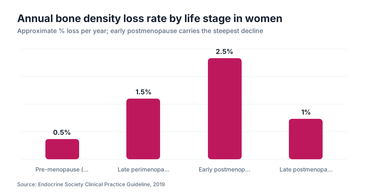
The steepest drop comes in the first 5 to 7 years after menopause. The Endocrine Society reports women can lose 2% to 3% of bone density per year during this stretch, which adds up fast [4]. After that the rate settles to roughly 1% to 1.5% per year, but the early damage is already banked. A woman who reaches menopause at 51 and loses 3% a year for six years has shed close to 18% of her bone density before anything hurts.
This is why the timing of menopause matters so much for your skeleton. Women who reach menopause age earlier, naturally or after surgery, have a longer stretch of estrogen deficiency ahead and a higher lifetime fracture risk.
Men lose bone too, slower and without the hormonal cliff. That gap is why osteoporosis is mostly a women's disease. Roughly 80% of Americans with osteoporosis are women [3].
What do T-score and Z-score numbers actually mean?
Your DEXA report gives you two scores. Most decisions ride on the T-score, but the Z-score matters in younger women. Here's how they compare.
| Score | What it compares to | Normal range | Action threshold | |---|---|---|---| | T-score | Peak bone mass of a 30-year-old | -1.0 to +1.0 | Treatment usually considered at -2.5 or below, or -2.0 with a fracture risk factor | | Z-score | Age-matched peers | -2.0 to +2.0 | Z below -2.0 suggests a secondary cause worth investigating |
A T-score of -1.0 to -2.5 is osteopenia. The word sounds scary, but osteopenia by itself does not mean you'll fracture. Your doctor should also run your FRAX score, a free tool from the University of Sheffield that combines your BMD with age, prior fractures, smoking, alcohol, and family history to estimate your 10-year fracture probability [5].
If your 10-year probability of a major osteoporotic fracture is 20% or higher, or your hip fracture probability is 3% or higher, most U.S. guidelines say consider medication even without a formal osteoporosis diagnosis [3].
For premenopausal and younger postmenopausal women, the Z-score tells you more. A Z below -2.0 means your bones are thinner than your peers', which sends your doctor hunting for a cause: vitamin D deficiency, celiac disease, hyperthyroidism, long-term steroids, or an eating disorder.
What causes low bone density in women?
Estrogen loss is the single biggest driver, but it's far from the only one. Secondary causes of low bone density are common and get missed all the time [4].
Things that speed up bone loss: long-term corticosteroids (even inhaled steroids at high doses), proton pump inhibitors taken for years, some antiepileptic drugs, and aromatase inhibitors used in breast cancer treatment. Celiac disease, inflammatory bowel disease, and hyperthyroidism all mess with calcium absorption or bone metabolism. Eating disorders wreck bone, partly through estrogen suppression and partly through outright nutritional deficits.
Lifestyle matters too. Smoking is independently tied to lower BMD and more fractures. Heavy drinking (more than two drinks a day, regularly) blocks bone formation. Long bed rest or a sedentary life strips away the mechanical loading that tells bone to stay dense.
A lifetime of low calcium, especially in adolescence when you're building peak bone mass, compounds everything. The NIH Office of Dietary Supplements recommends 1,000 mg of calcium daily for women 19 to 50 and 1,200 mg daily for women 51 and older [6]. Most American women don't hit those numbers from food alone.
Vitamin D is calcium's partner. Without enough of it, your gut can't absorb calcium properly no matter how much you eat. The RDA is 600 IU a day for adults up to 70 and 800 IU past 70, though many endocrinologists aim for serum 25-hydroxyvitamin D levels of 30 to 50 ng/mL and find patients need 1,500 to 2,000 IU daily to get there [6].
How does menopause affect bone density, and how fast?
The link between estrogen and bone is direct and well settled. Estrogen receptors sit on both osteoblasts and osteoclasts. Estrogen pushes bone building and holds back breakdown. When ovarian estrogen falls during perimenopause, the balance tips hard toward breakdown.
The transition itself counts. Bone loss can start in late perimenopause, before your final period, as estrogen swings downward. Women with heavy hot flashes and night sweats, symptoms that track the size of the estrogen drop, tend to lose more bone in the first years after menopause [4].
By about 10 years out, the loss slows a lot. But by then a woman may have lost 10% to 20% of her peak bone mass. The cumulative lifetime hit from menopause-related estrogen loss can reach 35% of trabecular bone (the spongy inner bone in the vertebrae and the ends of long bones) and 25% of cortical bone (the hard outer shell) [4]. Trabecular bone is more metabolically active, so it responds fastest to estrogen changes and shows up first as vertebral fractures.
Not every postmenopausal woman gets osteoporosis. Genetics, body weight, lifetime calcium, exercise history, and how long you had estrogen on board all shift the path. Heavier women tend to have higher bone density, partly because fat tissue turns androgens into estrogen (a little protection) and partly because more weight means more load on the skeleton. This is one place where being slender works against you.
Does hormone replacement therapy protect bone density?
Yes. This is one of the most clearly supported benefits of hormone replacement therapy in the medical literature.
The Women's Health Initiative trial, complicated legacy and all, showed clearly that estrogen-progestin therapy cut hip fractures by 33% and vertebral fractures by 34% versus placebo over a median 5.6 years of follow-up [7]. That's a large, real effect. The protection held whether women started with normal density or already had osteopenia.
The mechanism is plain. Restoring estrogen to premenopausal levels re-engages its brake on osteoclasts, slows breakdown, and lets bone formation catch up. Spine and hip BMD rise in most women within one to two years of starting.
For women who start within 10 years of menopause or before age 60, the Menopause Society (formerly NAMS) holds that the benefits of hormone therapy for symptoms, bone, and cardiovascular risk generally outweigh the risks in healthy women in this window [8]. The estrogen patch and other transdermal forms may carry a lower clotting risk than oral estrogen, which matters if you have cardiovascular risk factors.
Progesterone goes alongside estrogen in any woman who still has a uterus, to protect the uterine lining. Some data hint that micronized progesterone (Prometrium) may treat bone a little more kindly than synthetic progestins, but the evidence isn't strong enough to decide anything on its own.
HRT is not a forever drug for most women. Stop it and bone loss resumes. Women who lean on HRT for bone protection and then quit in their 60s may need to move to a bisphosphonate or another bone-specific agent.
Do GLP-1 medications affect bone density?
This area is still moving, and the data are thin. Women using semaglutide or tirzepatide for weight loss should know the bone tradeoff.
Rapid weight loss from any cause reduces load on the skeleton and comes with modest bone loss at the hip and spine. The STEP 1 trial of semaglutide showed a mean 14.9% body weight reduction over 68 weeks, versus 2.4% on placebo [9]. Extension data reported small reductions in hip BMD in the semaglutide group compared to placebo, though the differences were modest and their clinical meaning is still argued. The semaglutide for weight loss literature has no long-term fracture outcome data yet.
GLP-1 receptors do sit in bone tissue, and some animal and early human work hints these drugs may act directly on bone metabolism, maybe in a good way. But animal data don't translate cleanly, and the weight-loss unloading seems to outpace any direct benefit in the human studies we have.
Here's the practical part. If you're on a GLP-1 and dropping real weight, this is the moment to be deliberate about resistance exercise, enough protein, calcium, and vitamin D. If you're also postmenopausal, a baseline bone density test is reasonable to raise with your clinician. Platforms like WomenRx that handle GLP-1 prescriptions alongside hormone therapy can look at this intersection on purpose, which is worth asking about if you're on both.
On a semaglutide vs tirzepatide head-to-head, the evidence doesn't clearly separate their bone effects.
How is bone density tested, and when should you get a DEXA scan?
The standard test is dual-energy X-ray absorptiometry, or DEXA (sometimes DXA). It takes 10 to 15 minutes, uses very low radiation (about a tenth of a chest X-ray), and measures BMD at the spine and hip [1].
U.S. Preventive Services Task Force guidelines recommend routine DEXA screening for all women 65 and older [10]. Under 65, the recommendation is to screen if your 10-year fracture risk is equal to or greater than that of a 65-year-old white woman with no other risk factors, which works out to about a 9.3% 10-year risk. You can check yours with the online FRAX tool.
Those are floors, not ceilings. Many clinicians and the Menopause Society say postmenopausal women under 65 with risk factors should get a baseline DEXA sooner. Reasons to test early: menopause before 45, surgical menopause, low body weight (BMI under 20), a first-degree relative with a hip fracture, a history of an eating disorder, long-term steroids, or secondary causes like celiac disease or hyperthyroidism.
If you're already on treatment, the usual repeat interval is two years to check response. Scanning more often rarely helps, because DEXA's precision means real change takes time to rise above the measurement noise. For what the procedure feels like and how to read your results, see our full guide to the bone density test.
Can you actually build bone density back, or only slow the loss?
Honest answer: it depends on where you're starting and what tools you use.
For most postmenopausal women with osteopenia handled by lifestyle alone, the real goal is slowing or stopping loss, not rebuilding. Exercise and nutrition prevent further decline. They don't usually reverse it at a population level.
Bisphosphonates (alendronate, risedronate, zoledronic acid) work mainly by shutting down osteoclast activity, so they slow breakdown. They do produce modest BMD gains of about 3% to 8% at the spine and 1% to 5% at the hip over three years, and they cut vertebral fracture risk by roughly 40% to 50% and hip fracture risk by about 40% in high-risk patients [3]. But much of that gain is loss prevented, not fresh bone built.
The drugs that build new bone are a different class. Teriparatide (Forteo) and abaloparatide (Tymlos) are synthetic pieces of parathyroid hormone. They fire up osteoblasts directly. In trials, teriparatide raised lumbar spine BMD by roughly 9% to 13% over 18 to 24 months, far more than any antiresorptive [4]. They're saved for severe osteoporosis or people who fracture despite bisphosphonates, partly because they cost tens of thousands of dollars a year and were long capped at a two-year total treatment window, a limit since updated.
Romosozumab (Evenity) is a newer antibody that blocks sclerostin, raising bone formation and lowering breakdown at the same time. It produces large gains (about 13% at the spine over 12 months) but carries a boxed warning about cardiovascular risk in patients with a prior stroke or heart attack [4]. It's a monthly injection for 12 months, followed by an antiresorptive.
Estrogen therapy, as above, both slows loss and delivers modest gains, and it's the only option that also handles menopausal symptoms.
Which exercises actually increase bone density?
Not all exercise builds bone. Swimming and cycling are great for the heart but give bone almost nothing, because the water or the bike carries your weight.
Bone responds to load and impact. Osteoblasts feel the strain and lay down more bone matrix. The exercises with the strongest evidence for holding or raising BMD fall into two buckets.
Weight-bearing impact: walking, jogging, hiking, dancing, stair climbing, and jumping. Higher impact means bigger strain signals. A meta-analysis in Osteoporosis International found impact exercise significantly improved lumbar spine BMD in postmenopausal women, with effect sizes roughly twice those of non-impact aerobic exercise [11].
Resistance training: weights or bands. The push and pull on bone during strength work is a strong build signal. Progressive resistance training (loading up gradually) at the spine and hip holds density and adds modest gains, and it builds the muscle and balance that cut fall risk, which matters at least as much as raw density for preventing fractures.
Do both. Three to four sessions a week, mixing impact with resistance work aimed at the hips, spine, and legs, is a reasonable evidence-based target. The exact exercises matter less than showing up and adding load over years.
Yoga and tai chi are worth it for balance and fall prevention even though their direct BMD effect is small. If you've already fractured or have severe osteoporosis, high-impact work carries its own fracture risk and should be guided by a physical therapist.
What supplements actually help, and which ones are a waste of money?
Calcium and vitamin D are the base, and both are genuinely evidence-based. The details decide how well they work.
Calcium: aim for 1,200 mg a day for women over 50 from food plus supplements combined [6]. Dairy, fortified plant milks, canned salmon with bones, and leafy greens (kale, bok choy) are good sources. If you supplement, calcium carbonate is cheapest and absorbs best with food. Calcium citrate costs more but absorbs equally well with or without food, which helps if you take acid reducers or have low stomach acid. Mega-doses above 1,000 mg a day of supplemental calcium have raised cardiovascular concerns in some studies (not all); keeping each dose at or below 500 to 600 mg and getting the rest from food is a sensible line.
Vitamin D: most women with limited sun need a supplement to reach 30 to 50 ng/mL. D3 (cholecalciferol) raises serum levels better than D2 [6]. 1,000 to 2,000 IU daily is a common maintenance dose, but the right number depends on your baseline level, your weight (more body weight means more to distribute through), and how much sun you actually get.
Magnesium: it feeds bone metabolism and vitamin D activation. Deficiency is common, especially in women on diuretics or with gut disease. The evidence that supplementing raises BMD is thin, but correcting a real deficiency makes sense. 300 to 400 mg daily from food and supplements combined is reasonable.
Collagen, strontium, and most bone-branded multivitamins: the evidence for real BMD change is weak to nonexistent. Spending on them instead of the basics is a bad trade.
Vitamin K2: some data suggest K2 (menaquinone-7) helps steer calcium into bone rather than arteries, mostly in populations with low baseline K intake. The effect in Western diets with adequate K is uncertain. It's low-risk, but don't treat it as a standalone bone fix.
How do you prevent bone loss if you can't or won't take hormones?
Plenty of women have real reasons to avoid estrogen, or simply choose not to take it. The non-hormonal options are good.
Bisphosphonates are the most prescribed class. Alendronate (Fosamax) is a once-weekly pill and the usual first choice. Risedronate (Actonel) is similar. Zoledronic acid (Reclast) is a yearly IV infusion, which some women stick with more easily than weekly pills. Oral bisphosphonates can irritate the upper GI tract, so take them with a full glass of water and stay upright for 30 minutes. Rare risks across the class include osteonecrosis of the jaw (mostly in cancer patients on high-dose IV therapy) and atypical femur fractures (after very long use, usually past 5 years). Most guidelines suggest a drug holiday after 3 to 5 years in lower-risk patients [3].
Denosumab (Prolia) is an antibody given as a subcutaneous shot every 6 months. It cuts vertebral and hip fractures well and works in women with kidney disease where bisphosphonates can't be used. One thing you must know: unlike bisphosphonates, denosumab leaves no residual effect in bone. Stop it abruptly without moving to a bisphosphonate and bone loss rebounds fast, with multiple vertebral fractures reported [4]. Never stop denosumab without a transition plan from your doctor.
Raloxifene (Evista) is a selective estrogen receptor modulator (SERM). It acts like estrogen in bone but blocks it in breast tissue. It cuts vertebral fracture risk and may lower breast cancer risk, but it doesn't reduce hip fractures and carries a clot risk similar to estrogen's. It's a good fit for some women who also want breast cancer risk reduction.
Lifestyle stays essential next to any drug. Medication works best on a base of enough calcium, vitamin D, resistance exercise, fall prevention, and not smoking.
Frequently asked questions
What is the normal bone density for a woman by age?
Bone density peaks around age 25 to 30, then gradually declines. Clinically, 'normal' is a T-score above -1.0 on a DEXA scan at any age. In your 50s, average spine T-scores often fall between 0 and -1.0. By your 70s, averages drift toward -1.0 to -1.5. But averages aren't targets. Your fracture risk depends on your own T-score plus factors like age and fall history.
At what age should a woman get her first bone density test?
The USPSTF recommends routine screening for all women 65 and older. If you're under 65 and postmenopausal with risk factors, such as early menopause, low body weight, steroid use, or a parent who broke a hip, many clinicians want a baseline DEXA sooner. Use the FRAX calculator to estimate your fracture risk. If it clears the threshold, insurance usually covers testing before 65.
Can you improve bone density after 50?
Real rebuilding is possible with anabolic drugs like teriparatide or romosozumab, which build bone directly and can raise spine BMD by 9% to 13% over 1 to 2 years. For most women after 50, though, the realistic goal is slowing loss, not reversing it. Bisphosphonates, hormone therapy, resistance exercise, and enough calcium and vitamin D can halt decline and add modest gains of 3% to 8% at the spine.
How much calcium do postmenopausal women actually need?
The NIH recommends 1,200 mg a day total from food and supplements for women over 50. Get as much as you can from food first, since high-dose supplemental calcium above 1,000 mg a day in one hit has raised cardiovascular concerns in observational studies. If you supplement, split doses of 500 to 600 mg absorb best and keep the risk low.
Does losing weight hurt your bones?
Yes, to a degree. Dropping body weight reduces load on the skeleton, and load is a signal bone uses to stay dense. Bariatric surgery studies show real bone loss, and GLP-1 trials like STEP 1 show modest hip BMD reductions alongside rapid weight loss. Counter it with resistance exercise, enough calcium and vitamin D, and adequate protein if you're losing significant weight by any method.
Is osteopenia dangerous, or should I worry about it?
Osteopenia (T-score -1.0 to -2.5) is a risk factor, not a disease. Whether it's dangerous depends on your full picture. Your doctor should run your FRAX score, which folds in age, BMD, and other factors to estimate your 10-year fracture probability. Most women with osteopenia and a low FRAX don't need medication, just lifestyle steps and monitoring. Those with a FRAX above the treatment threshold do benefit from drug treatment.
Does hormone therapy stop bone loss after menopause?
Yes. The Women's Health Initiative trial found combined estrogen-progestin therapy cut hip fractures by 33% and vertebral fractures by 34% versus placebo. It works by restoring estrogen's brake on osteoclasts, slowing breakdown. Spine BMD typically rises 1% to 3% a year in the first few years. When HRT stops, bone loss resumes, so plan a transition to another agent with your doctor.
What foods are highest in calcium for bone health?
Dairy is densest: a cup of plain yogurt has roughly 300 to 400 mg, a cup of milk about 300 mg. Non-dairy options include fortified plant milks (often 300 mg per cup), canned sardines with bones (about 325 mg per 3 oz), canned salmon with bones (about 180 mg per 3 oz), firm tofu set with calcium sulfate (about 200 to 400 mg per half cup), and cooked kale or bok choy (about 90 to 160 mg per cup).
Does vitamin D deficiency cause osteoporosis?
Severe vitamin D deficiency causes osteomalacia, where bone matrix fails to mineralize, which is different from osteoporosis. Chronic mild-to-moderate insufficiency drives poor calcium absorption and secondary hyperparathyroidism, and that does speed bone loss over time. Correcting deficiency is essential for any bone treatment to work, since even bisphosphonates and hormone therapy fall short without good calcium absorption.
How accurate is a DEXA scan, and can results vary between machines?
DEXA is the standard for BMD measurement, with good precision: the coefficient of variation is roughly 1% to 2% at the spine and hip. But machines from different makers aren't interchangeable. To track BMD over time, use the same machine, or at least the same manufacturer at the same site. A change that looks meaningful may partly reflect machine differences rather than true biological change.
What is the density of bone compared to other body tissues?
Cortical bone, the hard outer shell, has a material density of about 1.8 to 2.0 g/cm³, the densest tissue in the body apart from tooth enamel. Trabecular bone, the spongy inner structure, runs much lower at around 0.1 to 1.0 g/cm³ depending on site and person. For comparison, muscle is about 1.06 g/cm³ and fat about 0.9 g/cm³. That's why bone sinks and fat floats.
Are there symptoms of low bone density before a fracture?
Usually none. Osteoporosis is called a silent disease because losing bone causes no pain and no obvious signs. Many women first find out after a fragility fracture: a break from a fall at standing height or less, or a vertebral compression fracture that shows up as back pain or lost height. That's exactly why screening DEXA scans matter. Waiting for symptoms means waiting for a break.
Can semaglutide or other GLP-1 drugs affect bone density?
The evidence is still emerging. The STEP 1 trial of semaglutide showed modest hip BMD reductions alongside significant weight loss, likely from reduced load on the skeleton. GLP-1 receptors are present in bone and may act directly, but current human data show no net protective effect. Women on GLP-1 medications who are also postmenopausal should prioritize resistance exercise, calcium, and vitamin D, and discuss baseline DEXA timing with their clinician.
How long does it take to see improvement in bone density with treatment?
With bisphosphonates or hormone therapy, clear BMD gains on DEXA usually take 1 to 2 years to rise above measurement noise. Fracture risk drops faster, within the first year, because these drugs also improve bone quality and geometry in ways DEXA doesn't fully capture. Anabolic agents like teriparatide produce larger, faster gains, with significant spine BMD increases visible at 12 to 18 months.
Sources
- National Institutes of Health, National Institute of Arthritis and Musculoskeletal and Skin Diseases: Osteoporosis overview
- Cummings SR et al., JAMA 1993; Study of Osteoporotic Fractures: hip fracture risk and BMD
- Bone Health & Osteoporosis Foundation (formerly National Osteoporosis Foundation): Clinician's Guide to Prevention and Treatment of Osteoporosis
- Endocrine Society Clinical Practice Guideline: Pharmacological Management of Osteoporosis in Postmenopausal Women (2019)
- University of Sheffield FRAX Fracture Risk Assessment Tool
- NIH Office of Dietary Supplements: Calcium Fact Sheet for Health Professionals
- Women's Health Initiative Investigators, JAMA 2002: Risks and benefits of estrogen plus progestin
- The Menopause Society (formerly NAMS): 2022 Hormone Therapy Position Statement
- Wilding JPH et al. (STEP 1), New England Journal of Medicine 2021: Once-Weekly Semaglutide in Adults with Overweight or Obesity
- U.S. Preventive Services Task Force: Osteoporosis to Prevent Fractures Screening (2018)
- Zhao R et al., Osteoporosis International 2015: Effects of resistance and aerobic exercise regimes on bone mineral density in postmenopausal women