Bone density test: what it is, how it works, and what your results mean
TL;DR: A bone density test, most often a DEXA scan, uses low-dose X-rays to measure how much mineral is packed into your bones. It takes 10 to 30 minutes, is painless, and produces a T-score that tells you whether your bone density is normal, low (osteopenia), or low enough to qualify as osteoporosis. Women should typically get their first test at menopause or earlier if they have risk factors.
What is a bone density test, and why does it matter for women?
A bone density test measures the mineral content of your bones, most often your hip and lumbar spine, to estimate how strong they are and how likely they are to break. The version almost everyone gets is dual-energy X-ray absorptiometry, called a DEXA or DXA scan. It is the measurement the World Health Organization used to define osteoporosis in the first place [1].
Bone strength matters more for women than most people realize. Women lose bone faster than men, and the drop speeds up sharply around menopause because estrogen normally holds back the cells that break bone down [2]. By her late 50s or 60s, a woman's bone mass may be 20 to 30 percent below its peak in her early 30s. That loss is not fixed and it responds to treatment, but you cannot manage what you have never measured.
A bone density test is also called a BMD test (bone mineral density test). Lab reports and insurance paperwork use both terms. They mean the same thing.
The reason to test early is simple. Osteoporosis has no symptoms until a bone breaks. A hip fracture in a woman over 65 carries a one-year mortality rate of roughly 20 percent [3]. Catching low bone density before that happens is the entire point of the test.
How is a bone density test done, step by step?
The procedure is genuinely simple. You lie on a padded table, fully clothed (no gown needed unless you are wearing metal zippers or underwire). A flat arm extends over you, and a detector panel sits beneath the table. The machine sends two low-energy X-ray beams through your body at different energies. Bone absorbs those beams differently than soft tissue does, and the machine calculates how much mineral is present from how much of each beam gets through [4].
The scanner passes over your lower spine and then repositions to image your hip, usually one hip at a time. Some protocols also scan the forearm, particularly when the hip or spine measurements are not reliable. The whole appointment runs 10 to 30 minutes from the time you lie down, and most of that is positioning. The actual scan time for each site is closer to two to five minutes.
No injection. No dye. No tunnel. Essentially no discomfort. The radiation dose is tiny: a standard DEXA delivers roughly 1 to 10 microsieverts, less than the natural background radiation you absorb on a cross-country flight [5]. Pregnant women are still told to skip it and reschedule, purely out of caution.
After the scan, a technologist processes the images and a radiologist or your ordering physician reads them. You usually get a written report within a few days, sometimes the same day.
A few things throw off accuracy: recent barium contrast from a GI study, calcium supplements taken within 24 hours of the scan, severe arthritis in the lumbar spine (which can falsely inflate spine scores), and prior vertebral fractures in the area being scanned. Tell the technologist about all of these before you lie down.
How is a bone density test done on a woman specifically?
The mechanics of the scan are the same for men and women. What differs is the reference population used to generate your score. Your results are compared to the bone density of a healthy young adult woman at her peak bone mass, not to the average for your current age [1].
For women, the two sites scanned almost always include the posterior-anterior lumbar spine (usually L1 through L4) and the proximal femur (hip), which covers the femoral neck and total hip. These are the sites that best predict fracture and the sites the WHO classification is built around.
Menopausal status changes how your results are read, not how the scan is done. The Endocrine Society describes estrogen deficiency, whether from natural menopause, surgical menopause, or premature ovarian insufficiency, as the primary driver of accelerated bone loss in women [2]. If you are perimenopausal, your provider may order a baseline test earlier than the standard age-65 mark.
Women who have had breast cancer and are on aromatase inhibitors (which suppress estrogen harder than menopause alone) are generally tested right at the start of that therapy and then annually, because bone loss in that setting can be fast [6].
The scan itself takes the same 10 to 30 minutes no matter who you are. The difference is in how your doctor uses the numbers.
What do bone density test results mean? Reading your T-score and Z-score
Your report will have two numbers: a T-score and a Z-score. Most people, and most media coverage, focus only on the T-score.
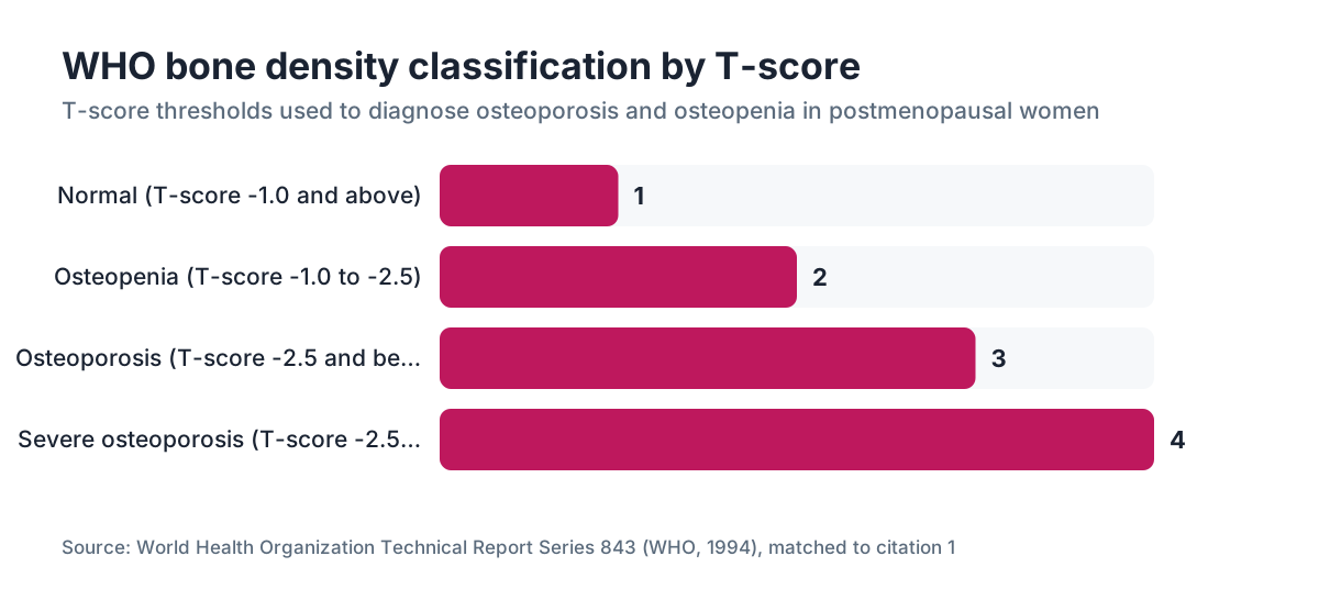
The T-score compares your bone mineral density to the average peak bone density of a healthy young adult of the same sex. The WHO classification is the worldwide standard [1]:
| T-score | Classification | |---|---| | -1.0 or above | Normal | | -1.0 to -2.5 | Osteopenia (low bone mass) | | -2.5 or below | Osteoporosis | | -2.5 or below with a fragility fracture | Severe (established) osteoporosis |
Each full point drop in T-score roughly doubles fracture risk, though the real relationship depends on which bone you are measuring and what other risk factors are in play.
The Z-score compares your bone density to other people your age and sex. A Z-score below -2.0 means your bone density is lower than expected for your age group and should trigger a workup for secondary causes: vitamin D deficiency, hyperparathyroidism, celiac disease, glucocorticoid use, or others.
One thing your T-score does not tell you on its own is your absolute fracture risk over the next 10 years. For that, clinicians use the FRAX tool, built at the University of Sheffield and endorsed by the WHO, which layers in age, sex, body weight, prior fractures, family history, smoking, alcohol use, glucocorticoid use, and secondary causes [7]. FRAX runs with or without a bone density score, though adding the femoral neck T-score sharpens it.
A FRAX result showing a 10-year major osteoporotic fracture risk above 20 percent, or hip fracture risk above 3 percent, is the threshold the Bone Health and Osteoporosis Foundation uses to recommend medication in the U.S. [3]. Your provider may use different thresholds depending on your full picture.
Do not panic if your T-score reads -1.5. Osteopenia is not a disease. It is a data point that tells you to pay attention to calcium, vitamin D, weight-bearing exercise, and, depending on your other risks, possibly medication or hormone therapy.
Who should get a bone density test and when?
The U.S. Preventive Services Task Force recommends bone density screening for all women 65 and older. For postmenopausal women under 65, the USPSTF recommends testing when a risk tool like FRAX puts your 10-year fracture risk at or above that of a 65-year-old white woman with no other risk factors, which works out to roughly a 9.3 percent 10-year major fracture risk [8].
That is the floor. In practice, plenty of clinicians test earlier and more often, especially with these risk factors:
- Early or surgical menopause (before age 45)
- Premature ovarian insufficiency
- More than three months of oral glucocorticoid (prednisone) use
- Aromatase inhibitor therapy for breast cancer
- A parent who fractured a hip
- Low body weight (BMI under 20)
- Smoking or heavy alcohol use
- Malabsorptive conditions (celiac, Crohn's, bariatric surgery)
- Rheumatoid arthritis or other inflammatory diseases
- Long-term use of proton pump inhibitors or anticonvulsants
If you are in perimenopause and carry two or more of these, it is entirely reasonable to ask your provider for a baseline DEXA now rather than waiting for 65. You can read more about when perimenopause typically starts at perimenopause age and what hormonal changes drive bone loss at menopause.
How often you repeat the test depends on where you start. Women with normal bone density may not need another scan for 10 to 15 years. Women with osteopenia are typically retested every 2 to 5 years. Women on osteoporosis medication are usually retested at 1 to 2 years to check whether the treatment is working.
How does menopause accelerate bone loss, and does hormone therapy help?
Estrogen is the main brake on osteoclasts, the cells that break bone down. When estrogen falls in menopause, osteoclast activity rises relative to osteoblast (bone-building) activity, and the net result is bone loss that can run 1 to 3 percent per year in the first few years after the final period [2].
The Endocrine Society's 2022 menopause guideline states that hormone therapy is effective for preventing bone loss and fractures and is FDA-approved for that use in postmenopausal women [9]. Menopausal hormone therapy, whether estrogen alone (for women without a uterus) or estrogen plus a progestogen, has preserved bone density in randomized trials, including the Women's Health Initiative.
This does not mean every woman with low bone density needs hormone therapy. That call weighs cardiovascular risk, breast cancer history, hot flashes, and personal preference. But if you are already thinking about hormone therapy for hot flashes or wrecked sleep, protecting bone is a real added benefit, not a footnote.
Progesterone's role in bone is murkier. Some data suggest a mild independent positive effect, but the evidence is nowhere near as strong as it is for estrogen. You can read the full picture at progesterone and hormone replacement therapy.
If you want a telehealth provider who can read your bone density results alongside your hormone levels and talk through MHT, WomenRx offers that kind of integrated hormone care for women in most U.S. states.
For women who cannot or choose not to use hormone therapy, FDA-approved osteoporosis medications include bisphosphonates (alendronate, risedronate, zoledronic acid), denosumab, raloxifene, and anabolic agents like teriparatide and romosozumab. The right choice depends on fracture risk, cost, tolerability, and kidney function.
Does GLP-1 weight loss affect bone density?
Yes, and it is an underappreciated question for women using semaglutide or tirzepatide. Rapid weight loss from any cause is tied to bone loss, because fat mass and muscle mass both load your bones mechanically, and when they drop fast, bone density can drop with them.
Data from the STEP trials of semaglutide showed modest reductions in bone mineral density, though how much that matters clinically is still being studied [10]. Tirzepatide data from the SURMOUNT program showed similar patterns.
The concern is not trivial, especially for women already approaching menopause and starting from a lower bone baseline. If you are on a GLP-1 and losing weight quickly, a baseline DEXA before or early in treatment is worth raising with your provider. You can read more on the bone side of things at semaglutide for weight loss and compare the two drugs at semaglutide vs tirzepatide.
Protecting bone during GLP-1 weight loss comes down to resistance training, enough protein (most data point to 1.2 to 1.6 grams per kilogram of body weight per day as protective), calcium, and vitamin D. These are not optional extras. They are the difference between losing fat and losing bone right alongside it.
What does a bone density test cost, and does insurance cover it?
For women 65 and older, Medicare Part B covers a DEXA scan once every 24 months with no cost-sharing [11]. Most private insurers follow the same guideline and cover the scan at 100 percent for women 65 and up.
Under 65, coverage depends on whether your insurer considers you high risk. If your physician documents specific risk factors, most major insurers will cover the scan, though you may owe a copay or have it count toward your deductible.
Without insurance, a DEXA scan at a hospital radiology department usually runs $150 to $350. Freestanding imaging centers and some health systems offer it for $75 to $150. The scan itself is cheap compared to most diagnostic imaging. The bigger question is whether you can get it covered.
Medicare covers a bone mass measurement for beneficiaries who meet one of five qualifying conditions, including estrogen deficiency with clinical risk factors for osteoporosis, verified by a physician [11].
If your provider orders the test, the CPT code for a DEXA scan of the axial skeleton (hip and spine) is 77080. For peripheral sites like the forearm, it is 77081. Knowing the code helps you check your benefits before you go.
What can you do to improve bone density after a bad result?
A low T-score is not a life sentence. Bone is living tissue and it responds to the signals you give it, though how much you recover depends on how low your score is, how old you are, and what caused the loss.
The interventions with the strongest evidence:
Resistance training is the most effective exercise for bone. Impact loading (jumping, running, hiking) helps too, especially at the hip. Yoga and swimming, excellent as they are for other reasons, do not load bone enough to move density in any real way. Aim for resistance training at least twice a week, hitting the major muscle groups.
Calcium from food beats supplements when you can manage it, both because food comes with co-nutrients and because high-dose calcium supplements (above 1,000 mg/day from pills alone) have been tied to a modest rise in cardiovascular events in some studies, though that data is contested. The National Academy of Medicine recommends 1,000 mg/day total calcium for women 19 to 50 and 1,200 mg/day for women over 50 [12].
Vitamin D is required for your gut to absorb calcium, so sufficiency is essential. Most guidelines recommend 600 to 800 IU daily for general adults, but many clinicians use 1,500 to 2,000 IU for women at risk of deficiency. A serum 25-OH vitamin D level above 30 ng/mL is generally considered enough for bone health [12].
Smoking speeds bone loss. Alcohol above two drinks a day does too. Neither is negotiable if bone is a priority.
For women with significant bone loss (T-score below -2.5, or osteopenia with a high FRAX score), lifestyle alone is usually not enough. FDA-approved medications can cut fracture risk meaningfully, sometimes by 40 to 70 percent for vertebral fractures with bisphosphonates [3]. That is a real number worth taking seriously.
Are there other ways to test bone density besides DEXA?
DEXA is the standard for good reasons: the most evidence, the best standardization, and WHO thresholds built around it. A few other technologies exist, but they play supporting roles.
Quantitative computed tomography (QCT) measures true volumetric bone density in three dimensions rather than the two-dimensional areal density DEXA gives you. It picks up trabecular bone changes more sensitively and shows up in research or specific clinical situations, but it delivers far more radiation and costs more, so it is not used for routine screening [4].
Peripheral DEXA (pDXA) scans the forearm, heel, or finger. It is cheaper and sometimes available right in a primary care office, but peripheral measurements cannot be swapped in for central (hip and spine) DEXA scores for diagnosis and treatment decisions.
Quantitative ultrasound of the heel shows up in some screening programs because it uses no radiation and travels well, but it is not diagnostic. A low heel ultrasound result should send you for a full DEXA, not replace one.
High-resolution peripheral quantitative CT (HR-pQCT) is a research tool that gives extraordinary detail about bone microarchitecture but is not available in routine clinical settings.
Here is the takeaway: if you need a diagnostic bone density test to make a clinical decision, insist on a central DEXA of the hip and spine. Peripheral and ultrasound tools can flag risk, but they cannot diagnose osteoporosis or guide medication.
How often should you repeat a bone density test?
The right interval depends on where you start. A large observational study in the New England Journal of Medicine found that women with normal bone density or mild osteopenia could safely wait about 15 years before rescanning, with little risk of slipping into osteoporosis in the meantime [13]. Women with moderate osteopenia should rescan in about 5 years; those with advanced osteopenia, about 1 to 2 years.
If you are on osteoporosis medication, most guidelines suggest a follow-up DEXA at 1 to 2 years to see whether bone density is responding. If you are stable on treatment after that, every 2 years is usually enough.
Women who stop hormone therapy should consider rescanning within 2 years, because bone loss can pick up speed once estrogen is withdrawn.
Medicare covers a scan every 24 months as a baseline, but your clinical situation may call for testing more or less often than that administrative default [11]. The interval belongs in a conversation with your provider, not a rigid calendar.
WomenRx providers can review your existing DEXA results next to your hormone panel and help you decide whether your current monitoring frequency fits your overall risk.
Frequently asked questions
What is a bone density test used for?
A bone density test measures the mineral content of your bones, mostly at the hip and spine, to estimate fracture risk. It is used to diagnose osteoporosis and osteopenia, track bone loss over time, guide treatment including medication and hormone therapy, and check response to that treatment. It is the standard screening tool for postmenopausal women.
How long does a bone density test take?
The full appointment is typically 10 to 30 minutes. The actual scanning time for each site, usually the lumbar spine and one hip, is closer to two to five minutes. Most of the appointment is setup and positioning. You stay clothed throughout and can go back to normal activity right after.
Does a bone density test hurt?
No. A DEXA scan is completely painless. You lie still on a padded table while a scanning arm passes over you. There is no injection, no dye, no compression, and no tunnel. The radiation dose is tiny, roughly comparable to a few hours of normal background radiation from the environment.
What is a normal bone density test result?
A T-score of -1.0 or above is classified as normal by the WHO. A T-score between -1.0 and -2.5 is osteopenia (low bone mass). A T-score of -2.5 or below is osteoporosis. Each full point drop in T-score roughly doubles fracture risk, though age, weight, and other factors matter significantly too.
At what age should a woman get her first bone density test?
The USPSTF recommends routine screening starting at age 65 for all women. Postmenopausal women under 65 should be screened if their 10-year fracture risk equals or exceeds that of a 65-year-old white woman with no other risk factors. Women with early menopause, long-term steroid use, or other major risk factors may benefit from testing in their 40s or 50s.
What is the difference between a T-score and a Z-score on bone density results?
A T-score compares your bone density to a healthy young adult of the same sex at peak bone mass. It is used to diagnose osteoporosis and osteopenia. A Z-score compares you to people your own age and sex. A Z-score below -2.0 suggests your bone loss is greater than expected for your age and should prompt a search for secondary causes like vitamin D deficiency or medication side effects.
Can you eat or drink before a bone density test?
Yes, you can eat and drink normally before a DEXA scan. The main prep instruction is to skip calcium supplements for at least 24 hours before the scan, since calcium residue in the GI tract can interfere with the image. Avoid barium contrast studies in the days before your scan, and tell the technologist about any metal hardware from prior surgeries.
Does menopause always cause bone loss?
Menopause consistently speeds bone loss because estrogen suppresses bone breakdown, and that brake comes off when estrogen falls. Most women lose 1 to 3 percent of bone mass per year in the first several years after their final period. Not every woman develops osteoporosis, but essentially all women lose some bone during and after the menopause transition.
Can hormone replacement therapy improve a low bone density test result?
Hormone therapy is FDA-approved for prevention of postmenopausal osteoporosis. It consistently preserves bone density and has reduced fracture risk in randomized trials including the Women's Health Initiative. It does not usually produce large T-score gains but can stabilize density and prevent further loss. Whether it is right for you depends on your full medical history.
Will losing weight on Ozempic or Wegovy affect my bone density?
Rapid weight loss from GLP-1 medications like semaglutide can lower bone mineral density, because losing body mass reduces the mechanical load on bone. STEP trial data showed modest reductions in BMD with semaglutide. Women using these medications, especially those already at risk for low bone density, should prioritize resistance training, enough protein, calcium, and vitamin D throughout treatment.
How accurate is a bone density test?
DEXA is highly reproducible on the same machine with trained technologists, with a measurement error of roughly 1 to 2 percent. The main sources of inaccuracy are patient positioning, prior vertebral fractures or severe arthritis (which can artificially inflate spine scores), body composition extremes, and comparing results across different machines. Always try to get follow-up scans at the same facility on the same machine.
Is osteopenia the same as osteoporosis?
No. Osteopenia (T-score between -1.0 and -2.5) means bone density is below average for a young adult but not low enough to qualify as osteoporosis. It does carry higher fracture risk than normal bone density, but most women with osteopenia do not go on to fracture without additional risk factors. Lifestyle changes are the primary intervention; medication may be added if the FRAX score is high.
What type of doctor orders a bone density test?
Primary care physicians, OB-GYNs, endocrinologists, rheumatologists, and internists all routinely order bone density tests. Any licensed provider with prescribing authority can order one. Telehealth providers can also order DEXA scans in most states, with the scan done at a local imaging center and results sent back to the ordering provider.
How is a DEXA scan different from a regular X-ray or MRI?
A regular X-ray shows bone structure but cannot accurately quantify bone density until 30 to 40 percent of bone mass is already gone. MRI does not measure bone mineral density at all. DEXA uses two X-ray beams at different energies to precisely calculate mineral content per unit area of bone. It is the only widely standardized method for diagnosing osteoporosis and tracking changes over time.
Sources
- World Health Organization, Assessment of fracture risk and its application to screening for postmenopausal osteoporosis (WHO Technical Report Series 843)
- Endocrine Society, Osteoporosis in Postmenopausal Women clinical practice guideline
- Bone Health and Osteoporosis Foundation (formerly National Osteoporosis Foundation), Clinician's Guide to Prevention and Treatment of Osteoporosis
- NIH National Institute of Arthritis and Musculoskeletal and Skin Diseases, Bone Density Scan (DEXA or DXA)
- FDA, Radiation-Emitting Products: Bone Density Scanning
- American Society of Clinical Oncology, Management of Osteoporosis in Survivors of Adult Cancers With Nonmetastatic Disease guideline
- FRAX Fracture Risk Assessment Tool, University of Sheffield / WHO Collaborating Centre
- U.S. Preventive Services Task Force, Osteoporosis to Prevent Fractures: Screening (2018)
- Endocrine Society, Treatment of Symptoms of the Menopause: An Endocrine Society Clinical Practice Guideline 2022
- Wilding JPH et al., STEP 1 Trial, New England Journal of Medicine 2021; 384:989-1002
- Centers for Medicare and Medicaid Services, Medicare Benefit Policy Manual, Chapter 15, Bone Mass Measurements
- National Academy of Medicine (formerly Institute of Medicine), Dietary Reference Intakes for Calcium and Vitamin D, 2011
- Gourlay ML et al., Bone-Density Testing Interval and Transition to Osteoporosis in Older Women, New England Journal of Medicine 2012; 366:225-233