Bone density scan procedure: what to expect, start to finish
TL;DR: A bone density scan (DEXA) measures bone mineral density at your hip and spine using low-dose X-ray. The procedure takes 10 to 30 minutes, uses no injections or enclosed tubes, and is painless. Medicare covers it every 24 months for eligible women. Results come as a T-score: -1.0 or above is normal, -1.0 to -2.5 is osteopenia, -2.5 or below is osteoporosis.
What is a bone density scan and why do women need one?
A bone density scan measures how much mineral is packed into a segment of bone. It is almost always done with dual-energy X-ray absorptiometry, which everyone shortens to DEXA or DXA. Denser bone is stronger bone. The scan is the clinical standard for diagnosing osteoporosis and osteopenia, two conditions that cause fractures and hit women far harder than men.[1]
Here is the reason women dominate those numbers. After menopause, estrogen falls sharply, and estrogen is one of the main signals telling bone-resorbing cells (osteoclasts) to slow down. Without that brake, women can lose 1 to 2 percent of bone mass per year in early menopause, sometimes more. The Bone Health and Osteoporosis Foundation estimates about 10 million Americans have osteoporosis, and roughly 80 percent of them are women.[2]
A DEXA does not diagnose fractures. It cannot tell you whether a bone is cracked right now. What it shows is bone mineral density (BMD) compared to two reference groups, which gives you two numbers: a T-score (versus a healthy young adult) and a Z-score (versus people your own age). The T-score is the one clinicians use to classify your bone health.
The scan also tracks how you respond to treatment. Start hormone therapy, a bisphosphonate, or another drug, and a follow-up DEXA in 1 to 2 years tells you whether it is working. Reading your bone density test results in that context is where the real value shows up.
Who should get a bone density scan?
The U.S. Preventive Services Task Force recommends routine screening for all women 65 and older.[3] Younger postmenopausal women should be screened when clinical risk factors are present. The tool most providers use to estimate that risk is FRAX, a fracture risk calculator developed by the World Health Organization.
Risk factors that pull the recommendation earlier:
- Early or surgical menopause (before age 45)
- Long-term glucocorticoid (steroid) use, even at low doses
- Rheumatoid arthritis
- Low body weight or a BMI under 18.5
- A parent who broke a hip
- Current smoking
- Heavy alcohol use
- Eating disorders or malabsorptive conditions like celiac disease
- Certain drugs: aromatase inhibitors, proton pump inhibitors, some antiepileptics
The North American Menopause Society adds that any woman who broke a bone as an adult from minimal trauma should be screened regardless of age.[4] "Minimal trauma" means a fall from standing height or less, the kind of stumble that healthy bone should shrug off.
Perimenopausal women nowhere near 65 often ask whether they should bother. Probably not yet, unless a risk factor above applies to you. Bone loss speeds up most in the first few years after your final period, so a baseline scan at or just after menopause age makes clinical sense. If you are in perimenopause with normal risk, ask your provider rather than ordering one on your own.
What does the bone density scan procedure actually involve?
The procedure is genuinely easy, which surprises most people. You lie on a padded table, fully clothed, though you will be asked to remove metal like belts and underwire bras. A scanner arm passes slowly over your body. You do not go inside a tube. No injection. No loud noise.
Standard clinical DEXA scans two sites: the lumbar spine (usually L1 through L4) and the proximal femur, meaning your hip. Some protocols add the non-dominant forearm, especially when the hip or spine cannot be measured well, say because of prior hip hardware or heavy arthritis in the spine.
Actual scanning runs 10 to 20 minutes for both sites. Total time in the building is closer to 30 to 45 minutes once you add check-in, positioning, and processing.
Radiation is very low. A spine-and-hip DEXA delivers roughly 1 to 6 microsieverts.[5] A standard chest X-ray delivers about 100 microsieverts, and a cross-country flight gives you around 10 microsieverts of background radiation just by sitting there. There is no meaningful radiation risk from DEXA.
Afterward, a radiologist or your ordering physician reads the scan. You typically get a report within a few days showing your T-scores, Z-scores, and sometimes a FRAX probability estimate.
How should you prepare for a bone density scan?
Preparation is minimal. A few things matter.
Skip calcium supplements for at least 24 hours before the scan. Calcium sitting in your GI tract can show up on the image and falsely inflate your apparent bone density. Most providers say your other medications are fine to take as usual.
Wear loose clothing without metal snaps, zippers, or underwire. Many facilities have you stash jewelry and belts in a locker.
Had a recent barium study, nuclear medicine scan, or contrast CT? Tell the scheduling desk. Leftover contrast can throw off DEXA results, and you may need to wait a few days to a week before your scan.
No fasting. No driver. You go back to normal life the second you leave.
For spine accuracy, tell your technologist about any spinal surgery, compression fractures, or severe arthritis. Bad degenerative disease at L1 through L4 can make the spine reading unreliable, because bone spurs and calcified discs look dense. When that happens, the hip result carries more weight in the interpretation.
If you are pregnant or think you might be, tell the facility first. DEXA is not routinely done during pregnancy, even though the dose is tiny.
How much does a bone density scan cost?
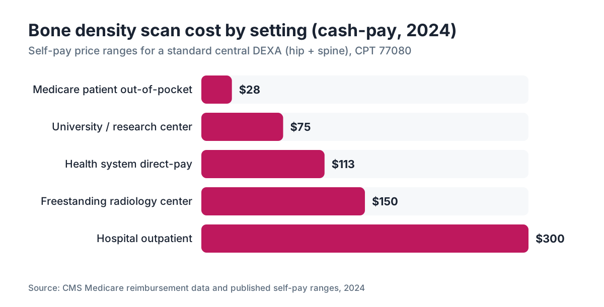
A bone density scan runs from about $28 out of pocket on Medicare to $400 cash at a hospital. Price swings on where you live, the facility type, and your insurance.[6]
Medicare Part B covers DEXA every 24 months (and more often if medically necessary) for qualifying beneficiaries. The Medicare-approved amount for a central DEXA (CPT code 77080) runs roughly $120 to $140 depending on your region, and you pay 20 percent after the Part B deductible, so out-of-pocket lands around $25 to $30 per scan.[6]
Most major commercial insurers cover DEXA for women 65 and older per USPSTF guidelines, and many cover it earlier for high-risk younger women. Policies differ, so verify before you schedule.
Cash-pay pricing ranges widely:
| Setting | Typical cash price | |---|---| | Hospital outpatient | $200-$400 | | Freestanding radiology center | $100-$200 | | Health system direct-pay program | $75-$150 | | University or research center | $50-$100 (sometimes subsidized) |
Uninsured, or your insurer denied the claim? Call the radiology center and ask for the self-pay rate. Radiology groups routinely knock 40 to 60 percent off the billed charge. Some community health centers offer DEXA free or on a sliding scale during osteoporosis awareness campaigns.
The scan itself is one of the cheaper diagnostic tests in women's health. The variable cost is really the physician visit needed to order it and read the results.
How do you read a T-score and what does it mean?
Your T-score is the number that sets your diagnosis. It compares your bone mineral density to the average peak density of a healthy young adult reference group, typically female, age 25 to 35.[1]
Here is the World Health Organization classification, which every DEXA report uses:
| T-score | Classification | |---|---| | -1.0 and above | Normal | | -1.0 to -2.5 | Osteopenia (low bone mass) | | -2.5 and below | Osteoporosis | | -2.5 and below with fragility fracture | Severe osteoporosis |
Each one-unit drop in T-score roughly doubles fracture risk, based on data from the Study of Osteoporotic Fractures.[1]
The Z-score is a different comparison. It measures you against age-matched peers. A Z-score of -2.0 or below means your bone loss runs ahead of what age alone explains, and it triggers a hunt for secondary causes: hyperparathyroidism, vitamin D deficiency, malabsorption, medication effects.
A few caveats worth carrying with you. The spine T-score can read falsely high because of arthritis, aortic calcification, or old vertebral fractures, especially in women over 60. Radiologists should flag this, but not all do. And T-scores from peripheral devices (wrist or heel ultrasound at a health fair) are not equal to central DEXA and cannot diagnose anything. They flag risk. They do not replace the real scan.
Your T-score is not your destiny. A T-score of -2.3 is no guarantee you will break a bone. Fed into FRAX alongside your age, prior fractures, and other factors, it becomes a 10-year probability. Treatment decisions run on that probability, not on the number by itself.
What happens if your bone density is low?
A result in the osteopenia or osteoporosis range starts a conversation, not an emergency. The first questions are root cause and fracture probability.
For osteopenia, first-line management is usually non-drug: weight-bearing exercise (both resistance training and walking stimulate bone), enough calcium (1,000 to 1,200 mg daily from food and supplements), vitamin D (800 to 1,000 IU daily for most postmenopausal women per the Endocrine Society), quitting smoking, and cutting back on alcohol.[7]
For osteoporosis, most guidelines add medication to those lifestyle steps. Oral bisphosphonates (alendronate, risedronate) are the most studied first-line drugs. Hormone therapy is FDA-approved to prevent postmenopausal osteoporosis, and the evidence that it reduces fracture risk is strong, which is one reason some providers favor it for women who also have menopausal symptoms.[4] An estrogen patch delivers estrogen through the skin and is one option inside hormone replacement therapy regimens with bone-protective data.
For high-risk women, anabolic drugs like teriparatide (Forteo) or romosozumab (Evenity) build new bone instead of just slowing loss. These are generally reserved for severe osteoporosis or women who fracture while already on a bisphosphonate.
If you are weighing hormone therapy partly for your bones, a telehealth provider like WomenRx can look at your full symptom picture and help you compare options without stacking up in-person referrals.
Follow-up scanning runs every 1 to 2 years if you are on treatment, or every 2 years if you are watching stable osteopenia without medication. Scanning more often rarely adds anything useful, and Medicare will not cover it more than every 24 months without documented medical necessity.
Does menopause affect your bone density scan results?
Yes. This is exactly where the hormone mechanism helps you read your report in context.
Estrogen suppresses osteoclast activity, the cells that resorb bone. When estrogen drops at menopause, those cells speed up and outpace the bone-building osteoblasts. The net result is bone loss, fastest in the first 3 to 5 years after your final period, then slower but steady.
The average postmenopausal woman loses about 1 to 3 percent of bone mineral density per year in early postmenopause without intervention.[4] Over a decade, that compounds. A woman entering menopause with a T-score of -0.5 (normal) can slide into the osteoporosis range by her mid-60s with nothing done.
Timing matters. Women with early menopause (before 45) have a longer stretch of estrogen deficiency and consistently show worse bone outcomes. NAMS treats premature ovarian insufficiency as an independent risk factor for osteoporosis and says these women should be considered for earlier screening, ahead of the USPSTF age line.[4]
Progesterone's part in bone is less settled than estrogen's. Some data suggest progesterone acts directly on osteoblasts, but the evidence that progesterone alone cuts fractures is not strong enough to steer treatment. Estrogen is the main bone-protective hormone in HRT.
For women just entering perimenopause, the practical read is this. Your scan at 50 or 55 is a snapshot of decades of estrogen exposure, diet, exercise, and genetics. It is a starting point, not a verdict.
How is a DEXA scan different from other bone health tests?
Several imaging and lab tests touch on bone health, and it helps to know which one your provider ordered and why.
Central DEXA (hip and spine) is the diagnostic standard. It is the only scan whose T-scores are validated against the WHO fracture threshold criteria.[1] When guidelines say "bone density scan," they mean central DEXA.
Peripheral DEXA measures the wrist or heel. Faster and cheaper, sometimes used for population screening. It cannot diagnose osteoporosis by WHO criteria and should not drive treatment decisions.
Quantitative CT of the spine gives volumetric BMD and can separate trabecular from cortical bone. More sensitive to early change, but it delivers more radiation and costs a lot more. Mostly research and specialized clinics.
Heel ultrasound (quantitative ultrasound) uses no radiation and costs little. It is often the device at health fairs. It tracks with fracture risk across populations but is no substitute for DEXA in an individual patient.
Lab tests that go with a low-density workup include serum calcium, phosphorus, 25-hydroxyvitamin D, PTH, TSH, and sometimes 24-hour urine calcium. These find secondary causes. They do not measure bone density itself.
Vertebral fracture assessment (VFA) is an add-on to DEXA that images your spine on the same machine to look for existing compression fractures. Plenty of postmenopausal women carry silent vertebral fractures that change their treatment category. NAMS recommends VFA for women 70 or older with a T-score below -1.0, or younger women with real height loss or back pain.[4]
Can semaglutide or GLP-1 medications affect bone density?
This question lands in providers' inboxes constantly right now. The honest answer: we do not have clean long-term data yet, but there are reasons to watch.
Rapid weight loss of any kind, including from GLP-1 receptor agonists like semaglutide, cuts the mechanical load on bone. Bone is load-sensitive. The more you weigh and the more impact force runs through your skeleton, the more bone your body keeps. Drop weight fast and bone density tends to follow.
Data from the STEP trials of semaglutide for weight management showed modest drops in total body BMD, on the order of 0.5 to 1 percent over 68 weeks, more than weight loss alone would predict in some analyses.[8] What that means clinically is still uncertain. Most endocrinologists prescribing GLP-1s for weight loss now suggest:
- Baseline DEXA before or shortly after starting, especially for women over 45
- Enough protein (0.8 to 1.2 g per kg body weight) to hold onto lean mass
- Resistance exercise to keep mechanical load on bone
- Vitamin D and calcium monitoring
If you are considering semaglutide for weight loss and you are peri- or postmenopausal, a baseline DEXA is a reasonable test to have on file, separate from your fracture-risk screening timeline. The WomenRx clinical team factors this into a pre-treatment evaluation when it fits.
The semaglutide vs tirzepatide question on bone is not settled either. The SURMOUNT trials of tirzepatide showed similar modest lean mass and bone density shifts alongside large weight loss. The pattern looks like a class effect of GLP-1-driven weight reduction, not a specific drug toxicity.
What does a DEXA report look like and what should you ask your doctor?
Your report lists T-scores and Z-scores for each site measured, usually as a number plus a color-coded chart (green for normal, yellow for osteopenia, red for osteoporosis). It may include a FRAX calculation showing your 10-year probability of a major osteoporotic fracture and of a hip fracture specifically.
The FRAX thresholds most U.S. guidelines use to recommend medication are a 10-year major osteoporotic fracture probability of 20 percent or higher, or a hip fracture probability of 3 percent or higher.[9] These are not hard rules, but they are the most cited clinical thresholds in the Bone Health and Osteoporosis Foundation guidelines.
Questions worth asking when you review results:
- Was the spine result usable, or did degenerative changes make it unreliable?
- What is my FRAX score, and does it change how you read my T-score?
- Do I have secondary causes of bone loss that labs should rule out?
- If my Z-score is low, what explains it beyond age?
- What rescan interval do you recommend for me?
- If I start treatment, what target T-score or FRAX change are we aiming for?
An osteopenia result (T-score -1.0 to -2.5) often stirs up anxiety way past the actual risk. A 52-year-old with a T-score of -1.5, no prior fractures, and no other risk factors may have a 10-year hip fracture probability under 1 percent. That is very low risk and does not call for medication. Flip it: a 68-year-old with a -2.3, a prior wrist fracture, and low body weight may sit at a 5 to 7 percent 10-year hip fracture risk, well past the threshold for a treatment discussion.
Frequently asked questions
How long does a bone density scan take?
The scanning itself takes 10 to 20 minutes for both the hip and spine. Total time in the facility, including check-in, positioning, and processing, is typically 30 to 45 minutes. You can return to normal activities immediately after. No recovery time is needed.
Is a bone density scan painful?
No. A DEXA scan is completely painless. You lie on a padded table while a scanner arm passes over your body. There are no injections, no loud noises, and no enclosed spaces. Most people say the hardest part is holding still for a few minutes while the machine sweeps across the hip.
At what age should women get their first bone density scan?
The USPSTF recommends routine screening starting at age 65 for all women. Younger postmenopausal women with risk factors, including early menopause before 45, long-term steroid use, low body weight, or a parent who broke a hip, should be screened earlier based on clinical judgment and their FRAX score.
How much does a bone density scan cost without insurance?
Cash-pay prices range from about $75 to $400 depending on the facility. Hospital outpatient centers charge the most, typically $200 to $400. Freestanding radiology centers and direct-pay health systems often charge $75 to $150. Always ask for the self-pay rate; radiology groups routinely discount 40 to 60 percent off the billed charge.
Does Medicare cover bone density scans?
Yes. Medicare Part B covers a central DEXA scan every 24 months for eligible beneficiaries, and more often if medically necessary. Your cost-sharing is 20 percent of the Medicare-approved amount after the Part B deductible, which typically works out to $25 to $30 per scan at 2024 payment rates.
What is a normal T-score for a woman?
A T-score of -1.0 or above is classified as normal bone density by WHO criteria. A score between -1.0 and -2.5 indicates osteopenia (low bone mass). A score of -2.5 or below is osteoporosis. Each one-unit drop in T-score roughly doubles fracture risk, per data from the Study of Osteoporotic Fractures.
Do I need to stop taking calcium before a bone density scan?
Yes. Skip calcium supplements for at least 24 hours before your scan. Calcium in your GI tract can appear on the image and artificially inflate your apparent bone density, making the result less accurate. Food sources of calcium are fine. Most other medications can be taken normally.
Can you have a DEXA scan if you have a metal hip implant?
A hip implant makes that hip unmeasurable by DEXA, so the technologist scans the opposite hip and the lumbar spine instead. If both hips are replaced, the distal radius (forearm) can serve as an alternate site. This is a known limitation and should be flagged when you schedule your scan.
How often should you repeat a bone density scan?
Medicare covers repeat DEXA every 24 months. For women on osteoporosis treatment, most guidelines recommend rescanning every 1 to 2 years to check treatment response. Women with normal bone density and low fracture risk may only need a repeat scan every 3 to 5 years. Your rescan interval should be individualized with your provider.
Can weight loss from GLP-1 medications affect bone density?
Yes. Rapid weight loss reduces mechanical load on the skeleton, and data from the STEP semaglutide trials showed modest decreases in bone mineral density over 68 weeks. The clinical significance is still being studied. Women over 45 starting GLP-1 therapy for weight loss should consider a baseline DEXA and discuss calcium, vitamin D, and resistance exercise with their provider.
What is the difference between a DEXA scan and a bone ultrasound?
A central DEXA of the hip and spine is the clinical standard and the only scan whose results meet WHO criteria for diagnosing osteoporosis. Heel ultrasound (quantitative ultrasound) is radiation-free and inexpensive but cannot diagnose osteoporosis in an individual patient. It can flag elevated risk but should not replace a DEXA for diagnosis or treatment decisions.
Does hormone replacement therapy improve bone density?
Yes. Estrogen therapy is FDA-approved to prevent postmenopausal osteoporosis and has consistent evidence for reducing fracture risk. NAMS guidelines recognize HRT as an appropriate bone-protective option, particularly for women who also have menopausal symptoms. Women on HRT still benefit from a baseline and follow-up DEXA to document their response.
What is a vertebral fracture assessment (VFA) and should I ask for it?
VFA is an add-on to DEXA that uses the same machine to image your spine for silent compression fractures, which many postmenopausal women have without knowing. NAMS recommends VFA for women 70 and older with a T-score below -1.0, or for younger women with significant height loss. Finding an existing fracture upgrades your fracture risk category and can change treatment recommendations.
Can a bone density scan detect all types of fractures?
No. A DEXA scan measures bone mineral density but does not show current fractures. It predicts fracture risk. Vertebral fracture assessment (VFA) can identify prior spine compression fractures, but for other fractures you need conventional X-rays or a CT scan. DEXA is a risk tool, not a fracture diagnostic.
Sources
- U.S. Preventive Services Task Force, Osteoporosis Screening Recommendation
- North American Menopause Society (NAMS), 2021 Position Statement on Osteoporosis
- RadiologyInfo.org (American College of Radiology / RSNA), Bone Densitometry (DEXA)
- Centers for Medicare and Medicaid Services, Medicare Coverage of Bone Mass Measurements
- Endocrine Society Clinical Practice Guidelines
- Wilding JPH et al., STEP 1 Trial, New England Journal of Medicine, 2021
- Bone Health and Osteoporosis Foundation, Clinician's Guide to Prevention and Treatment of Osteoporosis
- FDA, Drugs@FDA Approved Drug Products database
- International Society for Clinical Densitometry (ISCD), Official Positions