Bone density scan: what the DEXA test measures and why it matters
TL;DR: A DEXA scan (dual-energy X-ray absorptiometry) measures bone mineral density in your hip and spine. The result is a T-score: above -1.0 is normal, -1.0 to -2.5 is osteopenia, below -2.5 is osteoporosis. The U.S. Preventive Services Task Force recommends screening for all women 65 and older, and earlier for postmenopausal women with risk factors.
What is a bone density scan and what does it actually measure?
A bone density scan, almost always done with a technology called dual-energy X-ray absorptiometry (DEXA or DXA), measures how much calcium and other minerals are packed into a segment of bone. The denser the bone, the more mineral content, and the stronger it is. The test focuses on the lumbar spine and the hip (usually the femoral neck and total hip) because those are the sites most likely to fracture and the ones with the best predictive data.
The machine works by passing two low-energy X-ray beams through the bone and comparing how much each beam is absorbed. Bone absorbs more radiation than soft tissue, and denser bone absorbs more than porous bone. That difference gets translated into a number called bone mineral density (BMD), expressed as grams per square centimeter (g/cm²). [1]
The raw g/cm² number is meaningful to your radiologist, but most patients and clinicians care about the T-score, which is what gets reported on your results sheet. More on that in the section on reading your results.
Some facilities also scan the forearm (distal radius), especially if you're very obese, have had parathyroid disease, or can't position correctly for a hip or spine scan. The forearm is a secondary site and doesn't replace hip and spine data for most decisions.
The test is not an MRI and not a CT. It uses a tiny fraction of the radiation of a standard chest X-ray, roughly 1-10 microsieverts depending on the machine and protocol, compared to 100 microsieverts for a chest X-ray. [2] That's why it can be repeated every one to two years without meaningful radiation concern.
Who should get a DEXA bone density scan, and when?
The U.S. Preventive Services Task Force (USPSTF) recommends bone density screening for all women age 65 and older, and for postmenopausal women younger than 65 whose 10-year fracture risk is equal to or greater than that of a 65-year-old white woman with no additional risk factors (roughly 9.3% on the FRAX tool). [3]
The National Osteoporosis Foundation (now part of the Bone Health and Osteoporosis Foundation) and the Endocrine Society set similar thresholds and add several specific groups that should be tested earlier: postmenopausal women under 65 with a fracture from a minor injury, women stopping long-term glucocorticoids (prednisone and similar drugs), women with conditions known to cause bone loss like rheumatoid arthritis, celiac disease, or hyperthyroidism, and women who've lost more than 1.5 inches of height. [4]
Perimenopause is a window many women and even some clinicians miss. Bone loss speeds up in the two to three years before and after the final menstrual period, faster than at almost any other life stage. If you're in perimenopause with significant risk factors, asking for a baseline scan is reasonable even if you're 45 or 50. See perimenopause age for more on that timing.
Men get osteoporosis too, but this article focuses on women because the epidemiology is different: women lose bone faster after menopause and have lower peak bone mass to begin with. About 1 in 2 women over 50 will have an osteoporosis-related fracture in their lifetime. [4]
Insurance coverage under Medicare and most private plans follows the USPSTF recommendation, meaning a standard DEXA at 65 (or earlier with documented risk factors) is typically covered without out-of-pocket cost. Cash-pay DEXA scans run roughly $75 to $200 depending on facility and region, though some imaging centers charge less. [5]
How do you prepare for a DEXA bone density test?
Preparation for a bone mineral density DEXA scan is genuinely simple. Most people need to do almost nothing.
Stop calcium supplements 24 hours before the scan. Calcium tablets that haven't fully dissolved in your gut can sit on top of the area being scanned and artificially inflate your density reading. This is the one preparation step that actually matters clinically. Calcium from food is fine.
Wear comfortable, loose clothing without metal zippers, underwire bras, or thick metal buttons. Most facilities will let you keep your clothes on for a hip and spine scan if they're metal-free, though some will give you a gown anyway. Leave jewelry at home or be ready to remove it.
You do not need to fast. Eat and drink normally. The scan has no interaction with food.
If you've had a barium contrast study (like a barium swallow), a nuclear medicine bone scan, or any contrast-enhanced CT in the last seven to ten days, tell the technologist. Residual contrast material in your body can interfere with DEXA readings. You may need to reschedule. [6]
If you're pregnant or think you might be, tell them before the scan starts. The radiation dose is very low, but any X-ray during pregnancy is postponed unless medically urgent.
Bring a list of your medications, especially any that affect bone (bisphosphonates, steroids, aromatase inhibitors, thyroid hormone). This helps the reading radiologist and ordering physician interpret your results in context.
The scan itself takes 10 to 20 minutes from lying down to getting up. You lie flat on a padded table, usually without needing to hold still in an uncomfortable position. Most people find it considerably easier than an MRI.
How do you read your DEXA T-score and Z-score results?
Your DEXA report gives you two scores. The T-score and the Z-score. They answer different questions.
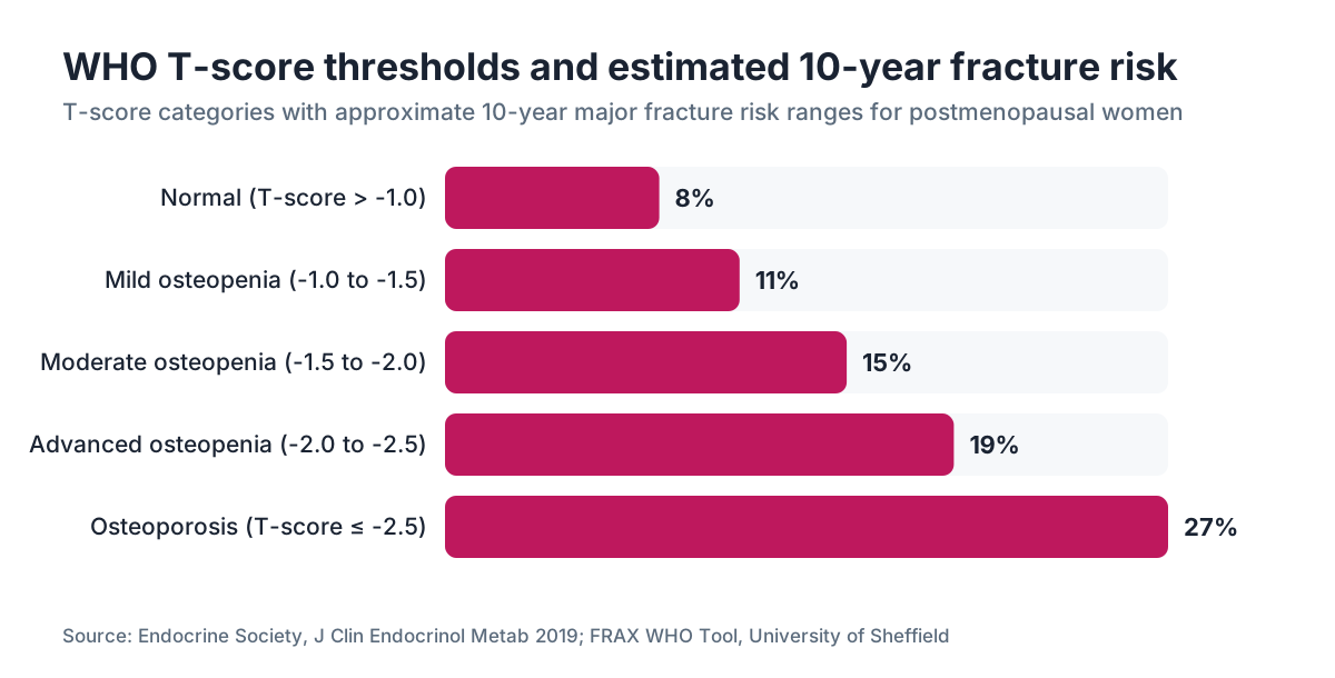
The T-score compares your bone mineral density to the average peak bone density of a healthy young adult (typically a 30-year-old of the same sex). The World Health Organization definitions, which are the ones clinicians actually use for diagnosis, are:
| T-score range | Diagnosis | |---|---| | Above -1.0 | Normal | | -1.0 to -2.5 | Osteopenia (low bone mass) | | -2.5 or below | Osteoporosis | | -2.5 or below plus a fragility fracture | Severe osteoporosis |
The Z-score compares your BMD to people of your own age, sex, and sometimes ethnicity. A Z-score below -2.0 means your bone density is lower than expected even for your age group, which should prompt a search for secondary causes of bone loss (like vitamin D deficiency, hyperparathyroidism, or medication effects) rather than just attributing everything to menopause. [1]
A few things to understand about T-scores that your result sheet probably won't explain. One point on the T-score scale corresponds to roughly a 10-15% increase in fracture risk per standard deviation decrease in BMD at the hip. [7] So going from -1.0 to -2.0 isn't just a number change; it represents a meaningful shift in your odds of a hip or spine fracture.
The T-score cutoffs were derived mostly from white postmenopausal women in the original WHO dataset. Their applicability to Black, Asian, and Hispanic women is imperfect. Black women tend to have higher bone density than white women at equivalent ages, which means a T-score of -2.5 may not carry the same fracture risk. Your clinician should interpret your results in context, more than hand you a printout.
Osteopenia is not a disease. It's a statistical category. Many women with T-scores between -1.0 and -2.5 will never fracture, especially if they have no other risk factors. A FRAX score (a free 10-year fracture probability calculator from the University of Sheffield) is the right next step when your T-score lands in osteopenia territory. [8]
What factors increase your risk of low bone density?
Bone density isn't just about calcium intake. It's the product of peak bone mass you built in your teens and twenties, the rate at which you're losing it now, and whether anything is accelerating that loss.
Estrogen is the single most important hormonal driver of bone maintenance in women. When estrogen drops in perimenopause and menopause, osteoclasts (bone-resorbing cells) become more active relative to osteoblasts (bone-building cells). The net result is bone loss that averages 1-2% per year in the early postmenopausal years, and can hit 3-5% per year in the first two to three years after the final period. [4] This is why menopause is the most common setting in which women first learn they have osteopenia.
Other major risk factors include:
- Low body weight or a BMI under 20 (less mechanical loading on bone)
- Smoking (impairs estrogen metabolism and bone formation)
- Excessive alcohol (more than 2-3 drinks per day inhibits osteoblast function)
- Family history of hip fracture
- Long-term use of glucocorticoids (prednisone 5 mg/day for more than 3 months)
- Aromatase inhibitors used for breast cancer treatment (suppress estrogen to very low levels)
- Hyperthyroidism or excessive thyroid hormone replacement
- Vitamin D deficiency (impairs calcium absorption)
- Prior low-trauma fracture after age 40
GLP-1 receptor agonists like semaglutide have attracted attention here because significant weight loss can reduce mechanical loading on bone. The STEP trials showed modest reductions in lean mass alongside fat mass, and some analyses suggest BMD at the hip may decline slightly with substantial weight loss. The FDA label for semaglutide notes that lean body mass loss accompanies overall weight loss. [9] This isn't a reason to avoid GLP-1 therapy, but it is a reason to monitor bone density and ensure adequate protein intake and resistance training if you're losing significant weight. You can read more about that tradeoff in our semaglutide for weight loss article.
How does menopause affect bone density and what does the evidence say about HRT?
Estrogen deficiency is the engine driving postmenopausal bone loss. This isn't controversial. The data on hormone replacement therapy and bone are among the most consistent in all of women's health.
The Women's Health Initiative (WHI) randomized trial found that combined estrogen-plus-progestin therapy reduced hip fracture risk by 34% and vertebral fracture risk by 34% compared to placebo. [10] Estrogen-only therapy in the WHI showed similar skeletal benefits. The Endocrine Society's guidelines on menopausal hormone therapy state that estrogen is effective for preventing bone loss and reducing fracture risk in menopausal women, and that for women who begin HRT near menopause, the benefits on bone (and cardiovascular health) are clearer than they are for women who start a decade or more after their final period. [4]
The practical implication: if you're in early menopause, have a T-score in the osteopenia range, and have no contraindications to estrogen, hormone replacement therapy is a legitimate first-line strategy for preserving bone. You don't have to go straight to bisphosphonates at a T-score of -2.0 if you're 50 years old and a good HRT candidate. That's a conversation worth having with your prescriber.
Progesterone has less clear evidence on bone directly, though it's included in combined HRT to protect the uterine lining and may have some bone-positive effects through progesterone receptors on osteoblasts. The data here are weaker than for estrogen.
For women considering or already using an estrogen patch, transdermal delivery achieves therapeutic estrogen levels with lower hepatic first-pass effects and is the preferred route for many women, particularly those with elevated cardiovascular risk factors. Bone-protective doses are well established for standard transdermal estradiol formulations.
For women who can't or won't use HRT, bisphosphonates (alendronate, risedronate, zoledronic acid) are the most studied pharmacologic alternatives. Denosumab, romosozumab, and teriparatide are options for more severe disease or women who don't tolerate bisphosphonates.
What's the difference between a DEXA scan and other bone tests?
DEXA is the reference standard for diagnosing osteoporosis and monitoring treatment. It's fast, low-radiation, widely available, and has decades of normative data behind it. That's why every major clinical guideline uses DEXA T-scores as the diagnostic criteria.
Peripheral DXA (pDXA) scans the wrist, finger, or heel. You sometimes see these in pharmacies or health fairs. They're inexpensive and convenient, but they can't diagnose osteoporosis by WHO criteria. They can flag someone as potentially at risk and prompt a referral for a full DEXA, but a heel scan alone isn't enough to make treatment decisions.
Quantitative CT (QCT) gives a true 3D measurement of volumetric bone density and can separate trabecular bone (the spongy inner structure) from cortical bone (the hard outer shell). It's more accurate in obese patients and gives additional structural information, but it uses more radiation, costs more, and has less normative data for fracture prediction. It's not a first-line test.
Quantitative ultrasound (QUS) measures bone density at the heel using sound waves. No radiation at all. But it's less standardized, can't be used to monitor treatment response, and isn't accepted for diagnosis.
VFA (vertebral fracture assessment) is an add-on to DEXA that images the spine to look for existing vertebral compression fractures. Many patients have these without knowing it. If your DEXA machine has this capability, it's worth asking for, especially if you've had back pain or visible height loss.
Serum markers like CTX (C-telopeptide of type I collagen) and P1NP (procollagen type I N-terminal propeptide) measure bone turnover, not density. They're useful for monitoring treatment response between DEXA scans, not for initial diagnosis.
How often should you repeat a DEXA scan?
Repeat scan timing depends on your baseline T-score, whether you're on bone-active treatment, and what clinical decisions would change.
For women with normal bone density who got screened at 65, re-testing every 15 years is often sufficient. A 2012 study in the New England Journal of Medicine followed 4,957 older women and found that women with normal baseline BMD took a median of 16.8 years to transition to osteoporosis. [7] Scanning every year when you have a normal T-score is almost never clinically necessary and is sometimes declined by insurance.
For women with osteopenia, the same study suggested rescreening intervals of 5 years for mild osteopenia (T-score -1.0 to -1.49), 3 years for moderate osteopenia (-1.5 to -1.99), and 1 year for advanced osteopenia (-2.0 to -2.49).
For women on treatment (bisphosphonates, denosumab, HRT), most guidelines suggest a repeat DEXA at 1-2 years to confirm treatment is working, then extending the interval if response is adequate. Medicare covers DEXA every 2 years for beneficiaries with documented osteoporosis or osteopenia.
If you're on denosumab specifically, the monitoring schedule is tighter because stopping that drug abruptly (without transitioning to a bisphosphonate) can cause rebound bone loss and vertebral fractures. Your prescriber should have a plan for this before you start.
At WomenRx, women starting or adjusting hormone therapy for menopausal symptoms often get a baseline DEXA as part of understanding their full bone health picture, which helps inform both the urgency of hormonal optimization and whether any additional therapy is needed.
What can improve bone density after a low DEXA result?
A low T-score is not a life sentence. Bone responds to mechanical loading, hormonal environment, and nutritional adequacy throughout life, even in your 70s.
Resistance training and weight-bearing exercise are the most consistently beneficial non-pharmacologic interventions. The mechanism is direct mechanical stress on bone, which stimulates osteoblast activity. Aim for at least two sessions of resistance training per week targeting the hip and spine. High-impact activity (jogging, jumping) adds additional benefit if your joints tolerate it. Swimming and cycling are great for cardiovascular health but provide minimal bone stimulus because they're low-impact.
Calcium and vitamin D are necessary but not sufficient. Most women get enough calcium from diet if they eat dairy, leafy greens, or fortified foods. The National Institutes of Health recommends 1,200 mg/day for women over 50 from all sources combined. Vitamin D targets are more debated: most experts aim for serum 25-hydroxyvitamin D above 20 ng/mL, with some endocrinologists preferring above 30 ng/mL for bone health. [11] Get your level checked before supplementing heavily; vitamin D toxicity is real at very high supplemental doses.
Protein intake matters more than most women realize. Bone matrix is about 30% protein, mostly collagen. Low protein intake is associated with worse outcomes after hip fracture and slower recovery. Aim for at least 1.0-1.2 grams of protein per kilogram of body weight per day if you're over 50.
For pharmacologic treatment, bisphosphonates (alendronate is generic and costs under $10/month) reduce vertebral fracture risk by about 40-50% and hip fracture risk by 20-40% in women with osteoporosis. [4] They're taken weekly (oral) or annually (IV zoledronic acid) and are the most prescribed first-line agents globally.
Estrogen therapy, as covered above, prevents bone loss effectively in women who start near menopause. For women whose primary reason for starting HRT is bone protection and who have no menopausal symptoms, the risk-benefit ratio requires more individual consideration; bisphosphonates may be the cleaner choice in that scenario.
Romosozumab and teriparatide are anabolic agents that actually build new bone rather than just slowing loss. They're reserved for severe osteoporosis (T-score below -2.5 with fracture, or -3.0 or lower) because of cost and administration requirements. Romosozumab carries a black-box warning for cardiovascular events in patients with prior heart attack or stroke. [12]
How much does a DEXA scan cost and is it covered by insurance?
Cost varies considerably depending on where you live, whether you have insurance, and what facility you use.
Under Medicare Part B, bone density measurements are covered every 24 months (or more frequently if medically necessary) for women with documented medical indications including estrogen deficiency, vertebral abnormalities, steroid use, hyperparathyroidism, or to monitor response to osteoporosis therapy. [5] Most beneficiaries pay 20% after the deductible; with a Medigap supplement, the out-of-pocket is often zero.
For women under 65 on private insurance, coverage follows the USPSTF recommendation (grade B), which means plans subject to the ACA must cover the test at 65 without cost-sharing, and many cover it earlier when a physician documents risk factors. Check your plan's explanation of benefits before scheduling.
Cash-pay prices range from about $75 at some independent imaging centers to $250 or more at hospital outpatient departments. Hospital outpatient pricing tends to be much higher for DEXA than the identical service at a freestanding imaging center. If you're paying out of pocket, call around. Some facilities offer self-pay discounts not advertised on their websites.
| Setting | Approximate cash-pay cost | |---|---| | Freestanding imaging center | $75-$150 | | Physician office (in-office DXA) | $100-$200 | | Hospital outpatient radiology | $150-$350+ | | Mobile DEXA services | $75-$125 |
Some bone health screening programs at universities and health fairs offer free or reduced-cost scans. The National Osteoporosis Foundation has periodically organized free screening events, particularly in May (Osteoporosis Awareness Month).
Can GLP-1 medications like semaglutide affect bone density?
This is a question women using semaglutide or tirzepatide for weight loss should ask, because rapid or substantial weight loss reduces mechanical loading on the skeleton. Bone remodels in response to load; when you weigh less, bones experience less compressive force and can lose density over time.
The STEP trials (the clinical program for semaglutide 2.4 mg) and the SURMOUNT trials (tirzepatide) did not show dramatic BMD reductions, but both programs were not primarily designed to assess bone outcomes, and most participants were on treatment for 68-72 weeks, which may not be long enough to see the full effect. [9] An analysis of STEP 1 data noted that while total fat mass decreased substantially, lean mass also declined, which matters for bone loading.
Major weight loss from any cause (bariatric surgery being the extreme example) is associated with increased fracture risk, particularly at the hip, and the effect is most pronounced when the weight loss exceeds 10-15% of body weight without concurrent resistance training.
The practical guidance: if you're using a GLP-1 agonist and losing significant weight, get a baseline DEXA if you haven't already, prioritize resistance training throughout your weight loss period, eat adequate protein (at least 1.0 g/kg body weight), and make sure your vitamin D and calcium are adequate. This isn't a reason to avoid these medications. It's a reason to manage bone health proactively alongside them.
See semaglutide vs tirzepatide for more on how those two GLP-1 options compare on body composition outcomes.
What happens after a bone density scan if your results are abnormal?
Getting a low T-score on paper is one thing. Knowing what to do next is another, and this is where a lot of women get lost, especially if they receive their results through an online portal without a follow-up appointment.
If your T-score is between -1.0 and -2.5 (osteopenia), the next step is almost always a FRAX calculation. Your clinician should enter your age, sex, weight, height, and clinical risk factors into the FRAX tool (available free from the University of Sheffield) to estimate your 10-year probability of a major osteoporotic fracture and hip fracture specifically. The treatment threshold that most U.S. guidelines use is a 10-year major fracture risk above 20% or a hip fracture risk above 3%. [8] Below those thresholds, lifestyle and monitoring are standard. Above them, medication is typically recommended.
If your T-score is -2.5 or lower, you meet the WHO diagnostic criteria for osteoporosis and treatment discussion is appropriate at that visit, not deferred.
A few labs should accompany the conversation: serum calcium, 25-hydroxyvitamin D, complete metabolic panel, thyroid function (TSH), and in some cases PTH, CBC, and serum protein electrophoresis to rule out secondary causes. Treating osteoporosis without checking whether something else is driving it is a mistake.
Fall prevention is part of bone health management that's easy to overlook. Balance training, medication review (sedatives, blood pressure drugs, and diuretics all increase fall risk), vision checks, and home hazard reduction are all meaningful. A hip fracture can occur at a T-score of -1.5 if the person falls; a person with a T-score of -3.0 who never falls has a lower absolute fracture rate than the data might suggest.
If you're working through hormonal optimization, weight management, or both, a telehealth platform like WomenRx can coordinate lab work, bone health monitoring, and treatment discussions in one place, particularly useful if you're managing menopause and a GLP-1 program simultaneously.
Frequently asked questions
What is a DEXA scan for bone density?
A DEXA (dual-energy X-ray absorptiometry) scan measures how much mineral is packed into your bones, most often at the hip and lumbar spine. The test uses two low-dose X-ray beams and takes 10-20 minutes. Results are reported as a T-score, which compares your bone mineral density to peak levels in a healthy young adult. It's the reference standard for diagnosing osteoporosis and osteopenia.
What T-score is considered normal on a DEXA bone density scan?
A T-score above -1.0 is considered normal. A T-score between -1.0 and -2.5 is classified as osteopenia (low bone mass). A T-score at or below -2.5 meets the World Health Organization diagnostic criteria for osteoporosis. These thresholds apply to postmenopausal women and men over 50; for younger premenopausal women, Z-scores are used instead.
How should I prepare for a bone mineral density DEXA scan?
Stop calcium supplements 24 hours before the scan because undissolved tablets can inflate your reading. Wear metal-free clothing. Eat and drink normally; no fasting required. Tell the technologist if you've had any contrast imaging (barium, nuclear medicine) in the past 7-10 days, as residual contrast can interfere with results. Bring a medication list. Pregnant women should postpone unless medically urgent.
At what age should women get their first bone density test?
The U.S. Preventive Services Task Force recommends screening at age 65 for all women, and earlier for postmenopausal women under 65 with a 10-year fracture risk equal to or greater than that of a 65-year-old white woman with no additional risk factors (roughly 9.3% on the FRAX calculator). Women with specific risk factors like steroid use, prior fragility fracture, or early menopause may benefit from earlier baseline testing.
Does hormone replacement therapy improve bone density?
Yes. The Women's Health Initiative trial found combined estrogen-progestin therapy reduced hip and vertebral fracture risk by 34% compared to placebo. Estrogen-only therapy showed similar bone benefits. The Endocrine Society supports HRT as an effective strategy for preventing bone loss in menopausal women, particularly those who start therapy near the time of menopause. Women with contraindications to estrogen have other pharmacologic options.
How is a DEXA scan different from a regular X-ray or CT scan?
A DEXA scan is specifically calibrated to measure bone mineral density and delivers about 1-10 microsieverts of radiation, far less than the 100 microsieverts from a chest X-ray. A standard X-ray doesn't quantify density, it only shows obvious structural changes. A CT scan gives more detailed 3D density data but uses much more radiation and costs more. DEXA is the clinical standard for osteoporosis diagnosis because of its precision and decades of comparative data.
Can semaglutide or other GLP-1 medications lower bone density?
Significant weight loss from any cause, including GLP-1 therapy, reduces mechanical loading on bone and may modestly lower BMD over time. The STEP trials for semaglutide 2.4 mg were not primarily designed to measure bone outcomes, but did show lean mass loss alongside fat loss. Women on GLP-1 medications losing more than 10% of body weight should prioritize resistance training, adequate protein, vitamin D, and consider baseline and follow-up DEXA scanning.
What is the Z-score on a DEXA report and how is it different from the T-score?
The T-score compares your bone density to peak young adult values and is used to diagnose osteoporosis. The Z-score compares your density to others of your own age, sex, and sometimes ethnicity. A Z-score below -2.0 means your bone density is lower than expected even for your age group, which suggests a secondary cause like vitamin D deficiency, hyperparathyroidism, or medication effects should be investigated rather than attributing the loss entirely to age.
How often should a bone density scan be repeated?
Repeat timing depends on your T-score. Women with normal bone density may only need rescreening every 15 years. Those with mild osteopenia can wait 5 years; moderate osteopenia, 3 years; advanced osteopenia, 1 year. Women on osteoporosis treatment typically repeat in 1-2 years to confirm response. Medicare covers DEXA every 24 months for eligible beneficiaries. Scanning more frequently than clinically indicated adds little useful information.
What exercises help increase bone density?
Resistance training (weights, resistance bands) and weight-bearing high-impact activities like jogging or jumping stimulate osteoblast activity and help maintain or modestly build bone. Aim for at least two resistance training sessions per week targeting the hip and spine. Swimming and cycling are good for cardiovascular health but provide minimal bone stimulus because the water and saddle absorb most mechanical load. Balance training reduces fall risk, which matters as much as density.
Does osteopenia always require medication?
No. Osteopenia is a statistical category, not a disease diagnosis. Many women with T-scores between -1.0 and -2.5 will never fracture. The standard next step is a FRAX calculation to estimate 10-year fracture probability. Most U.S. guidelines recommend treatment only when the 10-year major fracture risk exceeds 20% or hip fracture risk exceeds 3%. Below those thresholds, optimizing lifestyle, calcium, vitamin D, and hormone status is often the right approach.
Is a bone density scan covered by insurance?
Medicare covers DEXA every 24 months for women with documented risk factors, and most ACA-compliant private plans cover screening at age 65 without cost-sharing. Earlier coverage is often available with documented risk factors. Cash-pay prices range from about $75 at freestanding imaging centers to $350 or more at hospital outpatient departments. Calling an independent imaging center rather than a hospital radiology department usually gets you the lowest self-pay price.
What blood tests should accompany an abnormal DEXA result?
Before starting treatment for low bone density, check serum calcium, 25-hydroxyvitamin D, a complete metabolic panel, TSH (thyroid), and PTH (parathyroid hormone). In some cases, a CBC and serum protein electrophoresis (to rule out multiple myeloma) are appropriate. These tests identify secondary causes of bone loss that require their own treatment and can affect which bone therapy is safest.
Can perimenopause cause bone loss even before periods stop?
Yes. Estrogen levels begin fluctuating and declining in perimenopause, and bone resorption accelerates before the final menstrual period. Studies show bone loss can average 1-2% per year during perimenopause and reach 3-5% per year in the first 2-3 years after menopause ends. Women in perimenopause with risk factors can reasonably request a baseline DEXA to document their starting point before the most rapid phase of loss occurs.
Sources
- International Society for Clinical Densitometry, Official Positions 2019
- FDA, Radiation-Emitting Products: Bone Densitometry
- U.S. Preventive Services Task Force, Osteoporosis Screening Recommendation (2018)
- Endocrine Society, Pharmacological Management of Osteoporosis in Postmenopausal Women, J Clin Endocrinol Metab 2019
- Medicare.gov, Bone Mass Measurement Coverage
- Radiological Society of North America (RadiologyInfo.org), Bone Density Scan (DEXA)
- Gourlay ML et al., Bone-Density Testing Interval and Transition to Osteoporosis in Older Women, NEJM 2012;366:225-233
- FRAX WHO Fracture Risk Assessment Tool, University of Sheffield
- FDA, Wegovy (semaglutide) Prescribing Information
- Women's Health Initiative, Effects of Conjugated Equine Estrogen on Risk of Fractures, JAMA 2004
- National Institutes of Health, Office of Dietary Supplements: Vitamin D Fact Sheet for Health Professionals
- FDA, Evenity (romosozumab) Prescribing Information