Bone density loss in women: causes, timeline, and what actually helps
TL;DR: Women lose bone mass throughout life, but the steepest drop happens in the first 5-7 years after menopause, when falling estrogen can strip away up to 20% of bone density. Osteoporosis affects roughly 1 in 5 American women over 50. Diet, exercise, and hormone therapy all slow the loss. A DEXA scan tells you where you stand.
What is bone density loss and why does it matter for women?
Bone isn't static. Your skeleton is constantly being broken down by cells called osteoclasts and rebuilt by osteoblasts. When breakdown outpaces rebuilding, you lose bone mass. That net loss is what clinicians mean by bone density loss, or more formally, low bone mineral density (BMD).
For most women, bone mass peaks somewhere around age 25-30 and then slowly declines. The rate stays gentle through the 30s and early 40s. Then estrogen drops during perimenopause and menopause, and the osteoclasts win decisively. Estrogen normally puts a brake on bone resorption. Remove that brake and bone can disappear fast.
The clinical thresholds matter here. The World Health Organization defines osteoporosis as a DEXA T-score of -2.5 or below, and osteopenia (the warning zone before osteoporosis) as a T-score between -1.0 and -2.5 [1]. The National Osteoporosis Foundation estimates that about 10 million Americans have osteoporosis and another 44 million have low bone mass, and women account for roughly 80% of osteoporosis cases [2].
Why should you care if you feel fine? Because osteoporosis is a silent disease. You won't feel your bones thinning. The first sign is often a fracture, and hip fractures in older women carry a one-year mortality rate of 20-30% [3]. This isn't a cosmetic issue. It's a survival issue.
How much bone density do women lose during menopause?
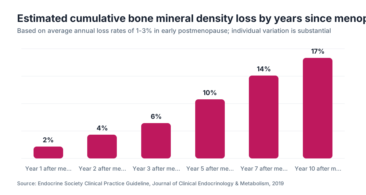
The numbers are sobering. In the years immediately surrounding the final menstrual period, women can lose 1-3% of bone mineral density per year [4]. Accumulated over the 5-7 years after menopause, that adds up to a possible 20% loss from peak bone mass. After that acute phase, the rate slows but never fully stops.
The Endocrine Society puts it plainly: "Estrogen deficiency is the dominant cause of bone loss in both women and men" [5]. That single fact explains why menopause is the biggest modifiable risk window for women's skeletal health.
A few things accelerate the loss beyond average. Surgical menopause (removal of both ovaries before natural menopause) causes an even sharper drop because estrogen falls overnight rather than gradually. Premature ovarian insufficiency (POI), which affects roughly 1% of women under 40, creates the same problem at a younger age, giving the skeleton decades of low-estrogen exposure before a woman even thinks about menopause [4].
Race and body size also matter. Asian and white women tend to start with lower peak bone mass and are at higher statistical risk than Black and Hispanic women, though no group is immune [2]. Thin women lose more because adipose tissue produces some estrogen after menopause, giving heavier women a small buffer.
What causes bone density loss beyond menopause?
Estrogen is the dominant driver, but it's not the only one. A useful mental model: anything that increases bone resorption, decreases bone formation, or impairs calcium absorption speeds up the loss.
Medication is a big category. Long-term glucocorticoids (prednisone, dexamethasone) are the most bone-toxic drugs in common use. Proton pump inhibitors (PPIs) reduce calcium absorption. Certain antidepressants (SSRIs), aromatase inhibitors used in breast cancer treatment, and depot medroxyprogesterone acetate (the injectable birth control) all have documented effects on BMD [3].
Lifestyle factors stack up quickly. Smoking is independently associated with lower bone density and higher fracture risk. Alcohol at three or more drinks per day suppresses osteoblasts. Extremely low body weight, seen in anorexia nervosa or during aggressive calorie restriction, starves the skeleton of the raw material it needs. Inadequate calcium and vitamin D intake is so common that the NIH estimates the majority of Americans fall short of recommended levels [6].
Secondary causes deserve a workup if your DEXA comes back worse than expected for your age. Hyperparathyroidism, hyperthyroidism, celiac disease, and chronic kidney disease all harm bone through different mechanisms. A good clinician will run labs before assuming your bone loss is purely menopause-related.
A note on GLP-1 receptor agonists: semaglutide and other semaglutide for weight loss drugs cause significant weight loss, and any rapid weight loss can reduce BMD because mechanical loading on bone decreases as body weight drops. The STEP 1 trial noted modest reductions in lean and bone mass with semaglutide [7]. If you're on a GLP-1 and losing weight fast, this is a conversation worth having with your prescriber, and a baseline DEXA is reasonable.
What are the symptoms of bone density loss?
Here's the frustrating truth: there are none. Bone density loss is clinically silent until you break something.
What you might eventually notice as osteoporosis advances are changes in height (losing more than 1.5 inches from your peak height is a red flag for vertebral compression fractures) or a rounding of the upper back called kyphosis. But by the time those signs appear, significant damage has already occurred.
Fractures that should make a clinician suspicious include wrist fractures from minor falls, vertebral fractures from sneezing or bending, and hip fractures from standing-height falls. These are called fragility fractures, and they are often the first diagnostic clue [3].
The bottom line: don't wait for symptoms. The whole point of bone density testing is catching low BMD before a fracture tells you about it.
How is bone density loss diagnosed?
The gold standard is a DEXA scan (dual-energy X-ray absorptiometry). It measures bone mineral density at the hip and lumbar spine, takes about 10-20 minutes, and exposes you to a tiny fraction of the radiation in a chest X-ray [1].
The result comes as a T-score (compared to a young adult reference population) and a Z-score (compared to age-matched peers). T-scores drive clinical decisions:
| T-score | Classification | |---|---| | -1.0 and above | Normal | | -1.0 to -2.5 | Osteopenia (low bone mass) | | -2.5 and below | Osteoporosis | | -2.5 and below with a fracture | Severe osteoporosis |
The U.S. Preventive Services Task Force recommends DEXA screening for all women 65 and older, and for younger postmenopausal women whose 10-year fracture risk is equal to or greater than that of a 65-year-old white woman [8]. That risk is calculated using the FRAX tool, which combines T-score with clinical factors like parental hip fracture history, smoking, and glucocorticoid use.
If you're in perimenopause with risk factors (early menopause, medication use, family history, low body weight), asking your doctor about an earlier baseline scan is reasonable. Medicare covers DEXA every 24 months for beneficiaries who meet criteria; private insurance coverage varies [8].
Does hormone replacement therapy actually prevent bone density loss?
Yes, and this is one of the best-supported findings in women's health research. Estrogen therapy preserves bone by suppressing osteoclast activity. The evidence goes back decades and has been replicated consistently.
The Women's Health Initiative (WHI) found that combined estrogen plus progestogen therapy reduced hip fracture risk by 34% and vertebral fracture risk by 34% compared to placebo [9]. That's a substantial effect. Estrogen-only therapy (for women without a uterus) showed similar skeletal benefits.
The North American Menopause Society (NAMS) states in its 2022 position statement that hormone therapy "is the most effective treatment for vasomotor symptoms and is effective for prevention of osteoporosis" in menopausal women [10]. That's about as clear an endorsement as clinical guidelines get.
Timing matters. The evidence is strongest when hormone therapy starts close to menopause onset, within the first 10 years or before age 60. Starting later doesn't appear to harm the skeleton, but the fracture-prevention data is thinner for late starters [10].
For women who can't or choose not to use hormones, there are good alternatives (discussed below). But if you're already considering hormone replacement therapy for hot flashes or mood, know that your bones are also benefiting. The skeletal protection is a real dividend.
Progesterone matters too, particularly for women with a uterus who need it to protect the uterine lining. Some data suggests micronized progesterone may have a mild additive benefit on bone compared to synthetic progestins, though estrogen is the primary driver of the skeletal effect.
Delivery method is less important than consistency. Whether you use an estrogen patch, gel, spray, or oral estradiol, the skeletal benefit depends on reaching adequate serum estradiol levels and maintaining them.
What non-hormone medications treat bone density loss?
For women who can't or won't use hormone therapy, and for those whose osteoporosis is severe enough to warrant additional pharmacotherapy, there are several well-studied drug classes.
Bisphosphonates are the first-line prescription option for most postmenopausal women with osteoporosis. Alendronate (Fosamax), risedronate (Actonel), and zoledronic acid (Reclast, given IV annually) all reduce osteoclast activity and cut fracture risk by roughly 40-50% at the spine and 20-40% at the hip [3]. Generic alendronate is inexpensive, often under $10 per month. The main complaints are GI side effects with oral forms and a small risk of atypical femur fracture with very long-term use (beyond 5-10 years), which is why drug holidays are sometimes recommended.
Denosumab (Prolia) is a biologic injection given every six months. It blocks a protein (RANK ligand) that drives osteoclast formation. It's effective but has a significant rebound risk: stopping denosumab without transitioning to a bisphosphonate can cause rapid, severe bone loss within 12-24 months [3].
Romosozumab (Evenity) is a newer anabolic agent that simultaneously builds bone and slows resorption. It's given monthly by injection for 12 months and then followed by antiresorptive therapy. It's generally reserved for severe osteoporosis or high fracture-risk patients because it carries a cardiovascular warning [3].
Raloxifene (Evista) is a SERM (selective estrogen receptor modulator) that protects bone and reduces breast cancer risk but can worsen hot flashes and carries a similar clot risk to estrogen. It's a reasonable option for women primarily concerned about vertebral fractures.
Calcitonin (Miacalcin nasal spray) is older, has modest fracture data, and has largely fallen out of favor in current guidelines [2].
How much calcium and vitamin D do you actually need?
The National Institutes of Health recommends 1,200 mg of calcium per day for women 51 and older, up from 1,000 mg for younger adults [6]. Vitamin D recommendations are set at 600-800 IU per day, though many bone specialists think that's too conservative for women with documented deficiency [11].
Food first is the right approach. Dairy, fortified plant milks, leafy greens, sardines with bones, and tofu set with calcium sulfate are all solid sources. A cup of milk provides roughly 300 mg; a serving of cooked kale provides about 100 mg. If you can get most of your calcium from food, you'll absorb it more efficiently than from supplements.
For supplements, calcium carbonate is cheapest but requires stomach acid for absorption (take it with food). Calcium citrate is better absorbed without food and is the better choice if you use PPIs or have low stomach acid. Splitting doses helps because absorption falls sharply above 500 mg at once [6].
Vitamin D status should ideally be checked with a blood 25-hydroxyvitamin D level. Most clinicians aim for 30-50 ng/mL [11]. Getting there often requires 1,000-2,000 IU of supplemental D3 daily, especially in northern latitudes during winter. Magnesium (320 mg/day for women) also supports bone metabolism and is frequently short in women eating typical Western diets, though the fracture data on magnesium supplementation is less definitive.
What types of exercise build bone density?
Not all exercise is equal for bone. Swimming and cycling are great for cardiovascular health but essentially weightless for the skeleton, so they don't stimulate bone formation much.
What works is mechanical loading: impact and resistance. The National Osteoporosis Foundation specifically recommends weight-bearing aerobic activity (walking, hiking, jogging, stair climbing, dancing) and resistance training with progressive overload [2]. The key word is progressive. Walking the same route at the same pace year after year will maintain fitness but probably won't add bone once your skeleton adapts to that load.
High-impact activity, where both feet leave the ground, generates the greatest osteogenic signal. Studies have shown that even brief bouts of jumping (10-20 hops per day) can improve hip BMD in premenopausal and early postmenopausal women, though high-impact work is not appropriate for women with established osteoporosis who are at fracture risk.
For women with low bone density, the best exercise prescription is usually progressive resistance training (working with a trainer initially is worth the cost) plus moderate-impact weight-bearing activity, plus balance and stability work to reduce fall risk. Falls are the proximate cause of most hip fractures. Cutting fall frequency is as important as improving BMD.
Balance training, tai chi, and yoga all reduce fall risk in older women, with tai chi having the strongest randomized trial data [2]. Aim for at least 150 minutes per week of moderate weight-bearing activity and two resistance training sessions.
How does perimenopause affect bone density, and when should you start worrying?
Bone loss actually begins before your last period. Studies tracking women through the menopausal transition show that BMD starts declining in the two to three years before menopause, during late perimenopause, coinciding with the most erratic estrogen fluctuations [4].
If you're in perimenopause and wondering when menopause starts for most women, the average age of the final menstrual period in the U.S. is 51, but perimenopause commonly begins in the mid-to-late 40s and can last 4-10 years. That means your bones may be under accelerating stress for a decade before you're officially postmenopausal.
For women who reach natural menopause early (before 45) or who experience surgical menopause, the urgency is higher. Years of low estrogen before age 50 compound over a lifetime of skeletal exposure.
The practical implication: if you're in your mid-40s and your periods are irregular, it's not too early to assess your bone health. Know your family history, check your vitamin D level, tidy up your diet and exercise, and have a frank conversation with your clinician about whether a baseline DEXA makes sense for you now rather than waiting until 65.
WomenRx offers telehealth evaluation for women moving through perimenopause and menopause, including discussion of hormone therapy and bone health, which can be a useful starting point if you don't have a specialist nearby.
Can you rebuild bone density once it's lost?
You can recover some of it, but completely reversing significant bone loss is not realistic. Bisphosphonates, estrogen therapy, and especially the anabolic agents (teriparatide, abaloparatide, romosozumab) can meaningfully increase T-scores over time. Romosozumab, for instance, increased spine BMD by about 13% and hip BMD by about 6% over 12 months in the FRAME trial [12]. That's real, clinically meaningful gain.
But rebuilding bone from a severe T-score of -3.0 back to normal is not something current treatments accomplish. The more honest framing is stabilization and improvement: slowing further loss, adding back modest amounts of bone, and reducing fracture risk even beyond what T-score changes alone would predict. Some medications reduce fracture risk to a greater degree than their BMD gains suggest, probably because they also improve bone quality and microarchitecture.
The women who do best are those who catch bone loss early (osteopenia rather than established osteoporosis), address lifestyle factors aggressively, and use the appropriate medical therapy consistently over years. Consistency matters enormously. Bisphosphonate benefits largely disappear if you stop taking them.
One honest caveat: the data on exercise-induced bone gain in postmenopausal women is real but modest. Exercise is essential for fall prevention and general health, and it can slow further loss, but it probably won't move your T-score much on its own if you're already in osteoporosis range. You'll likely need pharmacotherapy too.
What's the connection between GLP-1 drugs and bone density loss?
This is an evolving area and deserves an honest, careful answer. GLP-1 receptor agonists like semaglutide and tirzepatide (see semaglutide vs tirzepatide) produce large weight losses, sometimes 15-25% of body weight. Any significant weight reduction reduces the mechanical load on the skeleton, and the skeleton responds by losing some mass.
The STEP 1 trial, which studied semaglutide 2.4 mg weekly for 68 weeks, showed a reduction in lean body mass as well as fat mass [7]. Bone mineral density changes were not a primary endpoint in the STEP trials, but secondary analyses and later research have noted modest BMD reductions, particularly at the hip, in people losing substantial weight on GLP-1 therapy.
GLP-1 receptors are also expressed in bone tissue, and there's some research suggesting GLP-1 agonists may have direct effects on bone turnover, potentially favorable ones. The net effect in humans at therapeutic doses is still being worked out.
Practical advice for women on semaglutide or compounded semaglutide who are losing significant weight: keep calcium and vitamin D intake dialed in, prioritize resistance training to preserve lean mass and bone, and consider a baseline DEXA if you don't have one. Don't stop a medication that's improving your metabolic health because of theoretical bone concerns, but do manage the risk proactively. Your prescriber should be factoring this into ongoing care.
What are the best strategies to prevent bone density loss starting now?
Prevention is genuinely more effective than treatment here, and the actions that work are not complicated, just underused.
First, know your numbers. A bone density test before or shortly after menopause gives you a baseline to track. Without it, you're flying blind.
Second, get your calcium and vitamin D from food first, then supplement the gap. Most women over 50 eating an average American diet get 600-800 mg of calcium per day from food and need supplemental D3 in the 1,000-2,000 IU range, but your specific needs depend on your serum 25-OH vitamin D level [6].
Third, lift weights. Progressive resistance training two to three times per week is probably the single highest-yield lifestyle change for maintaining bone after menopause. You don't need a gym. Bodyweight squats, lunges, pushups, and resistance bands all count. The load needs to increase over time.
Fourth, consider hormone therapy seriously if you're in the early postmenopausal window and have no strong contraindications. The data on fracture risk reduction is real, and for most women under 60 or within 10 years of menopause, the benefit-risk balance favors therapy [10]. This deserves a real conversation with a clinician, not a decision made off 2002 WHI headlines that have been substantially reinterpreted since.
Fifth, identify and modify your risk factors. If you smoke, stopping helps bone (and everything else). If you're on chronic steroids, ask about bone protective strategies. If you take PPIs, consider whether you still need them.
If your DEXA already shows osteopenia or osteoporosis, this is the time to discuss prescription therapy with your doctor. Waiting for a fracture to happen is not a strategy. WomenRx can help women connect with clinicians who take bone health seriously as part of an overall hormonal health plan.
Frequently asked questions
At what age does bone density loss start in women?
Peak bone mass arrives around age 25-30. After that, loss is slow until perimenopause begins, typically in the mid-to-late 40s. The steepest decline happens in the 5-7 years after the final menstrual period, driven by the sharp drop in estrogen. Women with surgical menopause or premature ovarian insufficiency face this decline earlier.
What is a normal bone density T-score for a 50-year-old woman?
A T-score of -1.0 or above is considered normal. Between -1.0 and -2.5 is osteopenia (low bone mass), and -2.5 or below is osteoporosis. Many 50-year-old women have T-scores in the osteopenia range, which is not an emergency but does call for lifestyle optimization and closer monitoring. The FRAX tool combines your T-score with other risk factors to estimate 10-year fracture probability.
How long does it take to lose significant bone density during menopause?
Studies show women can lose 1-3% of bone mineral density per year in the years immediately before and after menopause. Over the full 5-7-year acute loss phase, total loss can reach 20% from peak. The rate slows after that but doesn't stop. This is why addressing bone health in perimenopause, not at age 65, is the smarter window.
Does calcium supplementation actually prevent osteoporosis?
It helps, but it's not sufficient on its own. Meeting daily calcium requirements (1,200 mg for women 51 and older) through food and supplements reduces bone loss but doesn't eliminate it. Calcium works best as part of a broader program that includes vitamin D adequacy, weight-bearing exercise, and for many women, hormone therapy or prescription antiresorptive medication. Excess calcium supplementation beyond needs carries potential cardiovascular risk.
Can you reverse osteoporosis naturally without medication?
Lifestyle changes alone rarely move someone from osteoporosis back to normal BMD. Exercise, calcium, and vitamin D can slow further loss and modestly improve bone mass, but reversing established osteoporosis typically requires medical treatment. Anabolic agents like romosozumab or teriparatide build the most bone; bisphosphonates stabilize and slowly improve T-scores. Work with a clinician to decide what's appropriate for your T-score and fracture risk.
Is estrogen therapy safe for preventing bone loss?
For most women under 60 or within 10 years of menopause onset with no history of breast cancer, blood clots, or certain cardiovascular conditions, the evidence supports hormone therapy for bone protection. The North American Menopause Society's 2022 position statement endorses hormone therapy as effective for osteoporosis prevention. The risks differ by age, health history, type of hormone, and delivery method; individualized discussion with a clinician is essential.
How does vitamin D deficiency cause bone density loss?
Vitamin D is required for calcium absorption in the gut. Without enough vitamin D, you absorb much less dietary calcium even if your intake is adequate. The body then pulls calcium from bone to maintain blood levels, speeding up bone resorption. Vitamin D deficiency also raises parathyroid hormone levels, which directly drives bone breakdown. Checking your serum 25-hydroxyvitamin D level is the only reliable way to know if you're deficient.
What exercises are bad for osteoporosis?
Women with established osteoporosis should avoid high-impact activities like jumping or running if their fracture risk is high, and exercises that require forward spinal flexion (crunches, touching toes with rounded back) because these load the vertebrae in ways that can cause compression fractures. High-contact sports carry obvious fracture risk. Swimming and cycling are safe for cardiovascular fitness but don't build bone. A physical therapist can design a safe, effective program.
Do GLP-1 drugs like semaglutide cause bone loss?
Rapid weight loss from any cause, including GLP-1 receptor agonists, reduces mechanical loading on bone and can lower BMD modestly. Secondary analyses from GLP-1 trials have noted modest reductions in hip BMD. GLP-1 receptors exist in bone tissue, and direct effects are still being studied. Women on these medications should optimize calcium and vitamin D intake, do resistance training, and consider a baseline DEXA scan, particularly if other bone loss risk factors are present.
What blood tests should I ask for to evaluate bone health?
Beyond a DEXA scan, useful labs include serum 25-hydroxyvitamin D (checking deficiency), calcium, parathyroid hormone (to rule out hyperparathyroidism), thyroid-stimulating hormone (hyperthyroidism speeds up bone loss), complete metabolic panel (kidney disease affects bone), and in some cases bone turnover markers like serum CTX or P1NP, which can show whether bone resorption is elevated before a DEXA changes. Discuss which are appropriate with your clinician based on your history.
How does smoking affect bone density?
Smoking independently lowers bone mineral density through multiple mechanisms: it reduces estrogen levels, impairs calcium absorption, and is directly toxic to osteoblasts. Smokers have significantly higher fracture risk than nonsmokers, even after adjusting for other factors. The damage is partially reversible with cessation. This isn't a minor lifestyle footnote; the skeletal impact of smoking is clinically meaningful and should factor into fracture risk discussions.
What is FRAX and how is it used in bone density assessment?
FRAX is the WHO's fracture risk assessment tool. It calculates your 10-year probability of a major osteoporotic fracture (hip, spine, forearm, shoulder) using T-score combined with clinical risk factors like age, sex, body weight, parental hip fracture, smoking, alcohol, glucocorticoid use, rheumatoid arthritis, and secondary causes of osteoporosis. U.S. guidelines generally recommend treatment when FRAX shows a 10-year hip fracture risk over 3% or major fracture risk over 20%.
How often should I get a DEXA scan after menopause?
The U.S. Preventive Services Task Force recommends screening at age 65 for all postmenopausal women, and earlier for younger postmenopausal women with equivalent fracture risk. Medicare covers repeat DEXA every 24 months for eligible beneficiaries. For women on treatment for osteoporosis, most clinicians repeat DEXA every 1-2 years to monitor response. If your baseline is normal and risk factors are low, every 5 years may be enough. Discuss the interval with your doctor based on your individual risk.
Can low progesterone affect bone density?
There is some evidence that progesterone, particularly natural micronized progesterone, may support bone formation through osteoblast activity. Low progesterone during anovulatory cycles in perimenopause may contribute to bone loss independent of estrogen. However, the primary hormonal driver of postmenopausal bone loss is estrogen deficiency. Progesterone is included in hormone therapy for uterine protection, with any bone benefit being an added, if modest, effect rather than the primary mechanism.
Sources
- National Osteoporosis Foundation (Bone Health and Osteoporosis Foundation), Clinician's Guide to Prevention and Treatment of Osteoporosis
- American Society for Bone and Mineral Research, Primer on the Metabolic Bone Diseases and Disorders of Mineral Metabolism
- Endocrine Society, Osteoporosis in Postmenopausal Women Clinical Practice Guideline, Journal of Clinical Endocrinology & Metabolism 2019
- Endocrine Society, Endocrine Facts and Figures: Osteoporosis
- NIH Office of Dietary Supplements, Calcium Fact Sheet for Health Professionals
- Wilding JPH et al., STEP 1 trial, New England Journal of Medicine 2021
- U.S. Preventive Services Task Force, Osteoporosis to Prevent Fractures Screening Recommendation, 2018
- Women's Health Initiative Investigators, JAMA 2002 and WHI follow-up publications
- North American Menopause Society, 2022 Hormone Therapy Position Statement
- NIH Office of Dietary Supplements, Vitamin D Fact Sheet for Health Professionals
- Cosman F et al., Romosozumab FRAME trial, New England Journal of Medicine 2016