Bone density exam: what women need to know before, during, and after
TL;DR: A bone density exam, almost always a DEXA scan, measures how strong your bones are by comparing them to a healthy young adult baseline. Most women get their first scan at 65, or earlier after early menopause, long-term steroid use, or other risk factors. The test takes under 30 minutes and uses about a tenth the radiation of a chest X-ray. Results come back as a T-score: normal, osteopenia, or osteoporosis.
What is a bone density exam and what does it actually measure?
A bone density exam measures the mineral packed into a given area of bone, reported as grams per square centimeter. More mineral means denser bone, and denser bone resists fracture. Think of it as a structural audit. Not whether your bones look broken, but whether they're likely to break under ordinary stress.
The standard method is dual-energy X-ray absorptiometry, almost always called DEXA or DXA. Two low-dose X-ray beams pass through the body at once, and software calculates how much each is absorbed by bone versus soft tissue. It's fast and painless, and it uses about one-tenth the radiation of a standard chest X-ray [1]. The machine usually scans the lumbar spine (L1 through L4) and the hip, because fractures at those two sites carry the highest risk of disability and death in older women.
Some facilities also scan the forearm. That's useful when the spine and hip readings are hard to interpret, say if severe arthritis in your lower back is inflating the spine number.
Other technologies exist. Quantitative ultrasound of the heel shows up in community screening because it uses no radiation, but it isn't recommended for diagnosis or for tracking treatment response [2]. Peripheral DEXA devices scan the wrist or heel and share the same limits. For anything past a rough screen, central DEXA at the spine and hip is the standard everyone measures against.
What do your T-score and Z-score mean?
Your DEXA report shows two numbers, a T-score and a Z-score, and they answer different questions. The T-score compares your bone against a healthy young adult; the Z-score compares you against people your own age.
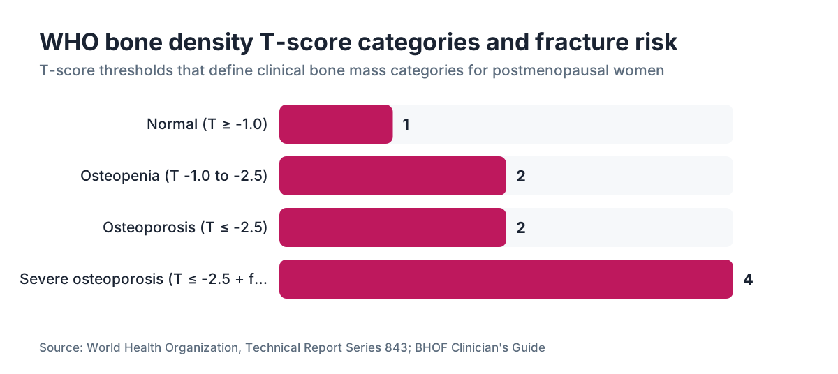
The T-score uses a reference database of healthy young adults at peak bone mass, typically women aged 20 to 29. The World Health Organization defines the categories this way [3]:
| T-score | Diagnosis | |---|---| | -1.0 and above | Normal bone mass | | -1.0 to -2.5 | Osteopenia (low bone mass) | | -2.5 and below | Osteoporosis | | -2.5 and below with a fragility fracture | Severe osteoporosis |
A T-score of -1.0 means your bone density sits one standard deviation below the young-adult average. Each full point below -1.0 roughly doubles fracture risk [4].
The Z-score compares you to women your own age and body size. A Z-score of -2.0 or lower in a premenopausal woman, or a man under 50, signals that something beyond normal aging is driving the bone loss and needs a workup for causes like hyperparathyroidism, malabsorption, or a medication effect. In postmenopausal women, T-scores drive the decisions and the Z-score is a secondary check.
Here's what providers often skip: DEXA measures areal bone mineral density, not true three-dimensional bone strength. Bone architecture, cortical thickness, and micro-fracture repair all shape fracture risk and none of them show up in a T-score. That gap is why FRAX, the fracture risk tool built by the WHO team at Sheffield, adds clinical risk factors on top of your T-score to produce a 10-year probability of major fracture and of hip fracture [4].
Who should get a bone density exam, and at what age?
The U.S. Preventive Services Task Force recommends routine DEXA screening for every woman aged 65 and older [5]. That's the headline. The earlier indications are where judgment actually matters.
For women under 65, the USPSTF recommends screening if your 10-year fracture risk equals or beats that of a 65-year-old white woman with no added risk factors, which FRAX puts at roughly 9.3% for any major osteoporotic fracture [5]. The Menopause Society and the Bone Health and Osteoporosis Foundation use the same framework and add specific triggers [2].
You should probably get a DEXA before 65 if any of these apply:
- You went through menopause before 45, whether spontaneous, surgical, or from chemotherapy. Estrogen loss is the single biggest driver of bone decline in women, and a longer estrogen-deficient stretch compounds the damage.
- You've taken corticosteroids (prednisone, dexamethasone) at 5 mg per day or more for longer than three months.
- A parent fractured a hip.
- You've had a fragility fracture, meaning a break from a fall at standing height or less.
- You have rheumatoid arthritis, celiac disease, inflammatory bowel disease, or another condition linked to bone loss.
- Your BMI is under 18.5.
- You smoke, or you drink more than two alcoholic drinks a day on most days.
Perimenopause alone doesn't earn you a scan. But if your periods are stopping early or you carry several risk factors, raise it with your provider instead of waiting until 65. Our piece on perimenopause age covers how the timing of menopause changes your bone trajectory.
How does menopause affect bone density and fracture risk?
Estrogen keeps bone loss in check by suppressing osteoclasts, the cells that break bone down. When estrogen falls in perimenopause and menopause, osteoclast activity surges while the bone-building osteoblasts can't match the pace. Women lose an average of 1 to 2% of spinal bone density per year in the first five years after the final period, and some lose 3 to 5% per year in that stretch [2]. The rate eases after those early years. It never fully stops.
Over a lifetime, women can lose 35 to 50% of trabecular bone (the spongy inner scaffolding, densest in the spine and hip) and 25 to 30% of cortical bone (the hard outer shell) [6]. Men lose bone too, just slower and without the sharp drop around a hormonal event.
By age 50, the Bone Health and Osteoporosis Foundation estimates one in two women will have an osteoporosis-related fracture in her remaining lifetime [6]. Hip fractures are the deadly ones. Roughly 20% of older adults who fracture a hip die within the following year from complications [6].
That's why menopause care and bone health can't be separated. Hormone therapy has decades of evidence that it holds bone density and cuts fracture risk when started around the time of menopause. The 2002 Women's Health Initiative found combined estrogen-progestin therapy reduced hip fractures by 34% and clinical vertebral fractures by 34% in postmenopausal women, though the WHI group was older (average age 63), so the absolute numbers need context for younger, recently menopausal women [7].
If you're weighing hormone replacement therapy or wondering whether an estrogen patch fits, bone protection is one of the honest reasons to have that talk sooner rather than later. Our overview of menopause walks through the wider picture.
How is a DEXA scan done, and what should you expect?
The test is anticlimactic in the best way. You lie flat on a padded table, fully clothed. Metal comes out of your pockets, but most scans skip the gown. A mechanical arm passes slowly over your lower spine, then your hip, usually one side. The scan itself runs about 10 to 20 minutes. Counting check-in and positioning, the appointment takes 30 to 45 minutes.
No injection. No contrast dye. The only prep is skipping calcium supplements the morning of the test, because extra calcium in your system can falsely raise the reading. Don't schedule a DEXA soon after a barium X-ray or a nuclear medicine scan, since leftover contrast interferes with the numbers.
Radiation dose is tiny, around 1 to 10 microsieverts depending on the machine and how many sites you scan. You absorb about 3,000 microsieverts a year from background radiation just living your life [1].
Results usually land within a few days. The report lists your BMD in g/cm², your T-score and Z-score for each site, and a short interpretation. Your ordering provider should walk you through the FRAX score alongside the raw numbers, because a T-score on its own won't tell you whether you need medication.
How often should you repeat a bone density exam?
Retesting runs on your result and your situation, not a fixed calendar. Normal bone at your first scan buys you years. Low bone means you check back sooner.
For postmenopausal women with normal bone density at baseline, the Bone Health and Osteoporosis Foundation notes that rescreening as far out as 15 years can be adequate if no new risk factors appear. Mild osteopenia (T-score around -1.5) can usually wait about 5 years. More advanced osteopenia (T-score -2.0 to -2.5) should be rechecked in 1 to 2 years [6].
Women on osteoporosis treatment (bisphosphonates, denosumab, and others) generally get a follow-up DEXA every 1 to 2 years to judge response. DEXA machines carry a built-in measurement variability of about 1 to 2%, so a change smaller than that may mean nothing. That threshold has a name, the least significant change. A good densitometry center tracks your scans on one machine to keep the comparison clean.
One practical rule: get every follow-up on the same machine at the same facility if you can. Machines from different manufacturers run slightly different reference databases, and switching between them can fake a change in your bone density that's really just calibration.
What can cause a false reading on a bone density exam?
DEXA is accurate but not bulletproof. Several things push your apparent density up or down.
Artifacts that raise apparent density: Severe lumbar osteoarthritis and degenerative disc disease are common in older women and lay down calcium in the joint spaces, making the spine read denser than it is. Vertebral compression fractures, which osteoporosis itself causes, can also inflate the AP spine T-score. Aortic calcification does the same. When your spine and hip scores split widely, or the spine number looks high for your overall health, an experienced radiologist should hunt for these.
Artifacts that lower apparent density: Metallic implants, spinal hardware, hip replacements, even calcium tablet fragments sitting in the bowel throw off the reading at that site. When that happens, the forearm or the opposite hip gets used instead.
Body composition: DEXA uses a two-compartment model, bone versus everything else, and very high fat mass can slightly underestimate true bone density. In heavier patients, the table weight limits (typically 300 to 450 lbs depending on the maker) can block the scan entirely.
Technician positioning: Rotating the femur during a hip scan changes the measured BMD. Quality control isn't optional.
What happens if your bone density is low, and what are your treatment options?
A low T-score is a starting point, not a verdict. Treatment turns on your 10-year fracture probability from FRAX, your age, your rate of loss, and any reversible causes.
For osteopenia without high fracture risk, lifestyle comes first. Calcium of 1,200 mg a day (food first, supplements only to close the gap) and vitamin D of 800 to 1,000 IU a day is a sane baseline, though megadoses add nothing and vitamin D toxicity is real above 4,000 IU per day [6]. Weight-bearing and resistance work (walking, jogging, dancing, lifting) tells bone to hold its density. Fall prevention matters enormously: balance training, clearing tripping hazards at home, reviewing any medication that makes you dizzy.
For osteoporosis, or osteopenia with a high FRAX score, medication is generally the call. The main options:
| Drug class | Examples | How they work | Typical use | |---|---|---|---| | Bisphosphonates | Alendronate, risedronate, zoledronic acid | Suppress osteoclast activity | First-line; oral weekly or IV yearly | | RANK-L inhibitor | Denosumab (Prolia) | Blocks osteoclast signaling | Injection every 6 months | | SERMs | Raloxifene (Evista) | Estrogen-like effect on bone | Postmenopausal women, also lowers breast cancer risk | | PTH analogs | Teriparatide (Forteo), abaloparatide (Tymlos) | Stimulate new bone formation | Severe osteoporosis or treatment failure | | Sclerostin inhibitor | Romosozumab (Evenity) | Builds bone and slows breakdown | Severe cases, capped at 1 year |
Hormone therapy is also an approved option for bone protection in women who have menopausal symptoms and are within 10 years of menopause or under 60. It isn't first-line purely for fracture prevention in symptom-free women, but for women who need it for hot flashes and broken sleep, the bone benefit is a real bonus reason to use it. If you're building a full hormone plan, our page on progesterone options is worth a read.
Women on GLP-1 medications like semaglutide for weight loss have an emerging concern worth knowing. Big weight loss of any kind, including from semaglutide for weight loss, takes mechanical load off your bones, and that load is part of what keeps density up. The STEP 1 trial recorded lean mass loss alongside fat loss, which matters for bone [11]. If you're dropping weight fast, a baseline DEXA plus adequate protein and calcium is sensible. WomenRx providers who prescribe semaglutide work bone health into that care, because losing weight without protecting muscle and bone is half a plan.
How much does a bone density exam cost, and does insurance cover it?
Medicare Part B covers a DEXA scan every 24 months for women who meet any of several criteria, including estrogen deficiency with clinical risk factors, vertebral abnormalities, long-term corticosteroid therapy, primary hyperparathyroidism, or monitoring of ongoing osteoporosis treatment [8]. After the deductible, the standard 20% coinsurance applies.
Private coverage is wide but not universal. The Affordable Care Act requires non-grandfathered plans to cover USPSTF A and B grade preventive services, which includes bone density screening for women 65 and older, with no cost-sharing [9]. For women under 65 scanning based on risk factors, coverage hangs on the specific plan and on whether the ordering provider documents the clinical reason.
Without insurance, a DEXA typically runs $125 to $250 at a hospital outpatient center, and sometimes less at independent radiology practices or health-system screening programs. Some academic medical centers run screening programs under $100. Prices swing a lot by region.
A vertebral fracture assessment (VFA), which can be added to the same DEXA visit, looks for compression fractures using a low-dose lateral spine image. It costs little extra and can catch silent fractures that would move you into a higher risk category.
How is a bone density exam different from a bone density test, and are there alternatives?
"Bone density exam" and "bone density test" mean the same thing in nearly every context. Both point to DEXA. If your paperwork or a referral says bone density test, you're getting the same procedure.
The main alternatives to central DEXA:
Quantitative CT (QCT) produces a true three-dimensional volumetric BMD and can separate trabecular from cortical bone. It gives more detail in some cases (complicated osteoarthritis, monitoring anabolic drugs) but delivers 10 to 100 times the radiation of DEXA and costs more. It's not a routine screen.
High-resolution peripheral quantitative CT (HR-pQCT) images the wrist or ankle with striking detail of bone microarchitecture. Right now it's mostly a research tool, available in a handful of centers.
Quantitative ultrasound of the heel is radiation-free and portable. A 2019 meta-analysis found acceptable sensitivity for flagging women at high fracture risk in large-scale screening, but current guidelines don't endorse it for diagnosis or for tracking treatment [2].
For most women, central DEXA is still the only practical, guideline-backed option for diagnosis and monitoring.
Can lifestyle and hormones actually improve your bone density after menopause?
Yes, though the ceiling sits lower than most people hope. Lifestyle changes reliably slow or stop further loss. They rarely rebuild much of what's already gone.
Resistance training is the strongest lifestyle lever. A 2022 meta-analysis in Osteoporosis International found progressive resistance exercise significantly raised lumbar spine and femoral neck BMD in postmenopausal women versus controls, with gains running 0.5% to 3% per year depending on intensity and duration [10]. That reads modest until you remember the untreated path is downhill.
Hormone therapy can genuinely add bone, more than hold the line. In the WHI, women on estrogen-alone therapy had 6.9% higher hip BMD and 5.2% higher spinal BMD after 3 years versus placebo [7]. The gains run larger when therapy starts in early menopause instead of a decade later.
Bisphosphonates raise spinal BMD by about 5 to 10% over 3 years and cut vertebral fracture risk by roughly 40 to 70% in trials, with hip fracture reductions near 40 to 50% for alendronate [6].
Anabolic agents (teriparatide, abaloparatide, romosozumab) build bone harder, with spinal BMD gains of 12 to 20% over 18 to 24 months in trials. They're saved for the most severe cases or for people who've plateaued on bisphosphonates.
The honest version: catch low bone mass early, in the osteopenia range, and lifestyle plus hormone therapy or a SERM where appropriate can keep you out of the fracture zone. Sit at -2.5 with a prior fracture and you almost certainly need medication. Calcium and walking won't carry that load alone.
Frequently asked questions
At what age should a woman get her first bone density exam?
The USPSTF recommends routine screening starting at age 65 for all women. Under 65, get one earlier if you went through menopause before 45, used long-term corticosteroids, have a parent who fractured a hip, or your FRAX 10-year fracture risk matches that of a 65-year-old woman with no added risk factors (roughly 9.3%). There's no reason to wait until 65 with clear risk factors on the table.
Is a bone density exam the same as a bone density scan?
Yes, the terms are interchangeable. Bone density exam, bone density scan, DEXA, and DXA all name the same dual-energy X-ray absorptiometry procedure. Some paperwork calls it bone densitometry. Unless your doctor specifically orders something different, like a quantitative CT, you're getting the same DEXA no matter which label appears.
Does a DEXA scan hurt or require any prep?
No, a DEXA scan doesn't hurt. You lie clothed on a table while a scanner arm passes over your lower spine and hip. The only real prep is skipping calcium supplements the day of the test and avoiding recent barium or nuclear medicine procedures. The whole appointment runs about 30 to 45 minutes from check-in to done.
What is a normal T-score for a woman in her 50s?
The WHO defines normal as a T-score of -1.0 and above. A 50-year-old in early menopause may already sit in the osteopenia range (-1.0 to -2.5), which isn't a disease but a signal to act on risk factors. T-scores in this decade swing widely with genetics, hormone history, exercise, and prior bone-affecting drugs. The trajectory and your FRAX-calculated 10-year fracture probability matter more than the single number.
Can perimenopause cause sudden bone loss?
Yes. The transition, when estrogen swings and then falls, is one of the fastest bone-loss windows in a woman's life. Spinal bone density can drop 1 to 5% per year in the first few years after the final period. That's why bone conversations belong in your 40s, not at 65. Early action, through lifestyle, hormone therapy, or medication, simply has more to work with.
Do GLP-1 drugs like semaglutide affect bone density?
Possibly. Big weight loss from any cause, including GLP-1 medications, takes mechanical load off your bones, and that load is one signal that keeps density up. The STEP 1 trial noted lean mass loss alongside fat loss, which matters for bone. Current data don't show dramatic BMD drops with GLP-1 use, but the research is young. Women on these drugs should keep calcium and protein adequate and consider a baseline DEXA if they carry other risk factors.
Does hormone replacement therapy actually prevent osteoporosis?
Yes. The Women's Health Initiative found combined estrogen-progestin therapy cut hip and clinical vertebral fractures each by 34% versus placebo. Estrogen-alone therapy showed similar or greater bone benefit. HRT works best for fracture prevention when started within 10 years of menopause or before age 60. It isn't first-line purely for bone in symptom-free women, but for women who also have menopausal symptoms, the bone benefit is a meaningful added reason.
What is the FRAX score and how does it relate to my DEXA results?
FRAX is a WHO-developed tool that calculates your 10-year probability of a major osteoporotic fracture (spine, hip, forearm, or shoulder) and of a hip fracture specifically. It combines your DEXA T-score with clinical risk factors: age, sex, BMI, prior fracture, parental hip fracture, smoking, alcohol, rheumatoid arthritis, and secondary causes. In the US, a FRAX above 20% for major fracture or above 3% for hip fracture generally triggers a medication recommendation.
How accurate is a DEXA scan, and can the results be wrong?
DEXA is the most accurate non-invasive method available, with real limits. Severe spinal arthritis, vertebral fractures, and aortic calcifications can falsely inflate spine readings. Metallic implants interfere with hip readings. Body size and positioning affect results. Precision runs about 1 to 2%, so small changes between scans may not be real. Follow-up scans on the same machine at the same facility give the most reliable comparison over time.
What medications can cause bone loss and increase fracture risk?
The most common culprit is long-term corticosteroid use (prednisone at 5 mg/day or more for 3 or more months). Others include high-dose proton pump inhibitors over years, selective serotonin reuptake inhibitors, anticonvulsants, some diabetes drugs (thiazolidinediones like pioglitazone), and aromatase inhibitors used in breast cancer. Depo-Provera (injectable medroxyprogesterone acetate) also reduces BMD, though some recovery happens after stopping.
Is osteopenia serious, and does it always progress to osteoporosis?
Osteopenia isn't a disease. It's a band of below-average bone density that raises fracture risk modestly. Not all osteopenia progresses to osteoporosis, and most osteoporotic fractures actually occur in women with osteopenia, simply because more women sit in that band. Whether it needs treatment depends on your full FRAX score, rate of loss, and other risk factors, not the T-score alone. Acting during the osteopenia stage genuinely makes a difference.
Can a bone density exam detect other bone conditions besides osteoporosis?
A standard DEXA is built to measure BMD and detect low bone mass. It's not a detailed diagnostic tool, but a radiologist reading the images may spot incidental findings like vertebral compression fractures, signs of Paget's disease, or obvious lesions. A vertebral fracture assessment (VFA) added to the session specifically screens for silent compression fractures. If bone cancer or metabolic bone disease beyond osteoporosis is suspected, MRI, CT, or a bone scan would follow.
How long does it take to get bone density exam results?
Results from a central DEXA usually arrive within 2 to 5 business days, though many facilities generate a preliminary report the same day for your ordering provider. A radiologist reads and signs the formal report. Your primary care provider, gynecologist, or endocrinologist then reviews it with you, ideally paired with your FRAX score and clinical history rather than just the bare T-score.
Should I get a bone density exam if I'm still premenopausal but have irregular periods?
Possibly, especially if the irregular periods reflect low estrogen (hypothalamic amenorrhea from undereating or overexercising, or premature ovarian insufficiency) rather than the high-estrogen cycling of PCOS. Low estrogen at any age speeds bone loss. If your periods have been absent or highly irregular for over a year and your estrogen is confirmed low, a DEXA is reasonable regardless of age. Ask your provider to read it by Z-score, which is how premenopausal results are interpreted.
Sources
- RadiologyInfo.org (American College of Radiology / Radiological Society of North America), Bone Densitometry (DEXA, DXA) patient page
- The Menopause Society (formerly NAMS), 2021 Position Statement on the Management of Osteoporosis in Postmenopausal Women
- World Health Organization, Assessment of Fracture Risk and Its Application to Screening for Postmenopausal Osteoporosis (Technical Report Series 843)
- FRAX Fracture Risk Assessment Tool, University of Sheffield
- U.S. Preventive Services Task Force, Osteoporosis to Prevent Fractures: Screening, 2018 Recommendation Statement
- Bone Health and Osteoporosis Foundation (formerly National Osteoporosis Foundation), Clinician's Guide to Prevention and Treatment of Osteoporosis
- Writing Group for the Women's Health Initiative Investigators, Risks and Benefits of Estrogen Plus Progestin in Healthy Postmenopausal Women, JAMA 2002
- Centers for Medicare and Medicaid Services / Medicare.gov, Bone Mass Measurements coverage
- HealthCare.gov, Preventive Care Benefits for Women
- Osteoporosis International (journal, Springer), 2022 meta-analysis of progressive resistance exercise and BMD in postmenopausal women
- Wilding JPH et al., Once-Weekly Semaglutide in Adults with Overweight or Obesity (STEP 1), New England Journal of Medicine 2021
- International Society for Clinical Densitometry, 2019 Official Positions (Adult)