Bone density DEXA scan: what women need to know
TL;DR: A DEXA scan (dual-energy X-ray absorptiometry) measures bone mineral density at the hip and spine in about 10 to 20 minutes. A T-score at or below -2.5 means osteoporosis; between -1.0 and -2.5 means osteopenia. The USPSTF recommends first screening at age 65, earlier if you have risk factors like menopause before 45 or long-term steroid use.
What is a DEXA scan and how does it measure bone density?
DEXA stands for dual-energy X-ray absorptiometry. Two X-ray beams at different energy levels pass through your bone, and the scanner calculates how much mineral is present by measuring how much each beam gets absorbed. The result is your bone mineral density (BMD), expressed in grams per square centimeter.
Axial DEXA, the clinical standard, scans the lumbar spine (L1-L4) and the proximal femur (hip). These are the two sites where fracture risk runs highest and where the numbers predict real-world outcomes best. Peripheral DEXA devices, which scan the wrist or heel, exist and cost less, but they are not the same test. The bone density test you want for fracture prediction is axial DEXA at hip and spine.
The radiation dose is tiny. The FDA puts a typical DEXA scan at roughly 1-10 microsieverts, on par with a few hours of background radiation from the environment [1]. A chest X-ray delivers about 100 microsieverts, ten times more. The scan itself takes 10 to 20 minutes, you stay clothed, and there is no injection or dye.
The machine outputs two comparison numbers: your T-score and your Z-score. The T-score compares your BMD to a young-adult reference population at peak bone mass. The Z-score compares you to people your own age and sex. Clinicians use T-scores to diagnose osteoporosis. Z-scores flag whether your bone loss runs faster than expected for your age, which points to a secondary cause worth chasing down.
What do T-scores and Z-scores actually mean?
The World Health Organization set the diagnostic thresholds that every clinician uses today [2]. The categories don't move:
| T-score | Diagnosis | |---|---| | -1.0 and above | Normal | | -1.0 to -2.5 | Osteopenia (low bone mass) | | -2.5 and below | Osteoporosis | | -2.5 and below with a fragility fracture | Severe osteoporosis |
Each standard deviation drop in T-score roughly doubles fracture risk. A woman with a T-score of -2.5 carries about twice the hip fracture risk of a woman at -1.5 [3].
Z-scores work differently. A Z-score below -2.0 counts as lower than expected for age and should trigger a workup for secondary causes: celiac disease, hyperparathyroidism, vitamin D deficiency, long-term glucocorticoid use, or hyperthyroidism. If your T-score is -2.8 but your Z-score is only -0.5, most of your bone loss comes from normal aging. If your Z-score is -2.3, something else is driving it.
One honest caveat. DEXA measures density, not architecture. Two women can share the same BMD and still have very different bone microstructure, and therefore different fracture risk. FRAX, the fracture risk assessment tool from the University of Sheffield, adds clinical risk factors on top of BMD to give a 10-year probability of major osteoporotic fracture [4]. Most treatment guidelines now pair FRAX with the T-score to decide who needs medication.
When should women get a DEXA scan?
The U.S. Preventive Services Task Force recommends routine DEXA screening for all women aged 65 and older [5]. For women under 65, the USPSTF recommends screening if their 10-year fracture risk equals or exceeds that of a 65-year-old white woman with no additional risk factors, which works out to about a 9.3% FRAX score for major fracture.
The Bone Health and Osteoporosis Foundation (formerly the National Osteoporosis Foundation) adds a longer list of reasons to scan earlier [6]. Get a baseline DEXA before 65 if any of these apply:
- Menopause before age 45, or surgical removal of ovaries
- More than three months of oral corticosteroid use
- A parent who had a hip fracture
- Low body weight (BMI under 20)
- Smoking or heavy alcohol use
- Rheumatoid arthritis
- A fragility fracture (breaking a bone from a fall at standing height or less)
- Conditions that impair calcium or vitamin D absorption
Perimenopause by itself is not a formal indication for early screening in most guidelines. But bone loss speeds up sharply in the two to three years before and after the final period. If you want to know where you stand before that window opens, a baseline scan in your late 40s or early 50s is a reasonable choice that most providers will support. See more on perimenopause age and when does menopause start to understand that timing.
If your first scan is normal and you have no major risk factors, repeat screening is typically every 15 years for T-scores between -1.0 and -1.5, every 5 years for scores around -1.5 to -2.0, and every 1-2 years for scores approaching -2.5 [3].
How does menopause affect bone density, and how fast does bone loss happen?
Estrogen is the main regulator of bone remodeling in women. It holds osteoclast activity in check, slowing the cells that break bone down. When estrogen drops at menopause, osteoclasts run relatively overactive and bone gets lost faster than it gets replaced.
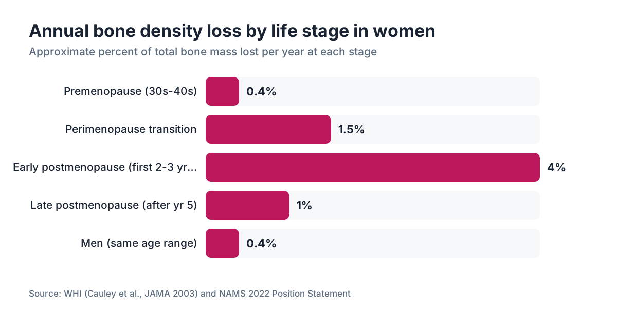
The numbers are stark. Women lose roughly 1-2% of bone mass per year during the menopausal transition, and up to 3-5% per year in the first two to three years after the final period [7]. Over a decade, that compounds to 15-25% of total bone mass. Men lose about 0.3-0.5% per year across the same age range.
The spine loses density faster than the hip in early postmenopause because trabecular (spongy) bone, which makes up most of the vertebral bodies, turns over more quickly than cortical (dense outer) bone. That is why spinal compression fractures often show up as the first sign of trouble.
By age 80, a woman's lifetime fracture risk sits at roughly 50%, on par with the combined risk of breast, uterine, and ovarian cancer [6]. That number isn't meant to scare you. It frames why bone density monitoring counts as clinical medicine, not optional wellness screening.
Learn more about hormone replacement therapy and its effect on bone, or read the estrogen patch overview for specifics on transdermal delivery and bone outcomes.
Does hormone replacement therapy actually improve DEXA results?
Yes, and this is one of the steadiest findings in menopause research. Estrogen therapy preserves and modestly increases bone mineral density at both hip and spine.
The Women's Health Initiative (WHI) showed that combined estrogen-progestogen therapy cut hip fracture risk by 34% and total fractures by 24% compared to placebo [7]. That result came from the full WHI cohort, not a pre-selected high-risk group. Estrogen-only therapy in women who had a hysterectomy showed similar fracture protection.
The North American Menopause Society (NAMS) 2022 position statement says hormone therapy is approved for the prevention of osteoporosis in postmenopausal women and is appropriate for women under 60 or within 10 years of menopause onset who have no contraindications [8]. NAMS sets no minimum fracture risk threshold for this indication, unlike bisphosphonate guidelines.
Progesterone enters the picture too. Some data, still short of definitive, suggests progesterone (the bioidentical form, not all synthetic progestins) has an independent bone-protective effect through progesterone receptors on osteoblasts. The clinical weight of that effect stays debated.
WomenRx providers review DEXA results alongside hormone levels when building HRT regimens, which is one reason having the scan number in hand before starting or adjusting therapy earns its keep.
If your provider is comfortable with your overall risk profile, starting HRT before a T-score falls into osteoporosis range is a legitimate preventive strategy, not an overreach.
How do GLP-1 medications affect bone density?
This is an open question in the field, and the honest answer is that we do not have long-term DEXA data from the major GLP-1 trials yet.
Here is what we know. GLP-1 receptor agonists like semaglutide and tirzepatide cause large weight loss, and rapid weight loss from any cause ties to bone loss. Fat mass and muscle mass both load bone mechanically; lose either and you cut that load. The STEP trials for semaglutide and the SURMOUNT trials for tirzepatide were not built to measure fracture endpoints, and BMD was not a primary outcome [9].
A 2023 meta-analysis in JBMR Plus found GLP-1 receptor agonists linked to small reductions in bone turnover markers, which could be protective, but the meaning of marker changes without fracture data stays limited [9]. In older adults with type 2 diabetes, semaglutide has not shown higher fracture rates in available data. Those patients aren't the same population as women taking semaglutide for weight loss who may already be perimenopausal.
Practically: if you are on a GLP-1 for weight loss and you are perimenopausal or postmenopausal, get a baseline DEXA before or shortly after starting. Keep calcium adequate (1,000-1,200 mg/day from diet plus supplements) and vitamin D (800-1,000 IU/day minimum), and put resistance training first. Resistance training has the strongest evidence for offsetting weight-loss-related bone loss. Semaglutide vs tirzepatide compares these two agents more fully, but from a bone standpoint neither holds a clear advantage in current data.
What factors affect the accuracy and reliability of a DEXA scan?
DEXA is reliable, but several things can skew results in either direction.
Artifacts that falsely raise bone density readings include vertebral compression fractures (calcified debris looks like dense bone), severe degenerative disc disease with osteophytes, aortic calcification, and dense surgical hardware in the scan field. This matters at the bedside. A 70-year-old woman with a reported spine T-score of -0.5 and visible arthritis on her imaging may have real osteoporosis hidden by artifacts. In that case the hip score carries more weight, or a quantitative CT (QCT) of the spine may replace it.
Factors that lower measured density include body position changes, different software versions between scans, and scanning on different machines. DEXA results are not perfectly interchangeable between manufacturers. Follow-up scans should happen on the same machine, or at least the same manufacturer's equipment, using a standardized protocol. The International Society for Clinical Densitometry (ISCD) publishes position statements on proper technique and quality control [10].
Body size and composition matter too. Larger individuals tend to show higher measured BMD, partly from geometry (bigger bones scatter more photons). Obesity does not protect against poor bone quality even when DEXA looks reassuring.
Insurance typically covers axial DEXA under specific diagnosis codes. Medicare covers one DEXA every 24 months for beneficiaries who meet criteria, including all women 65 and older [1]. Most commercial insurers follow similar logic but may require a qualifying diagnosis or risk factor for coverage before age 65. Cash pay prices run about $100 to $300 depending on facility and region.
What is the difference between a DEXA scan and other bone tests?
Several other imaging methods assess bone, and knowing what each one tells you (and what it can't) helps you have a sharper conversation with your provider.
| Test | What it measures | Fracture prediction | Cost estimate | Notes | |---|---|---|---|---| | Axial DEXA (hip + spine) | BMD at high-fracture-risk sites | Best validated | $100-$300 cash | Clinical standard | | Peripheral DEXA (wrist, heel) | BMD at peripheral sites | Moderate | $30-$75 | OK for initial screening; cannot guide treatment | | Quantitative CT (QCT) | Volumetric BMD; 3D | Strong, especially spine | $300-$600 | Better for artifact-prone spines; more radiation | | Trabecular bone score (TBS) | Bone microarchitecture from DEXA image | Adds to DEXA prediction | Add-on to DEXA cost | Useful in high-risk patients | | FRAX (no imaging) | 10-year fracture probability | Good population estimate | Free online | Uses clinical factors; BMD optional | | Heel ultrasound | Bone stiffness | Moderate | $20-$50 | Not diagnostic; research and community screening tool |
The trabecular bone score earns a note here. It is an add-on analysis run on the same DEXA image that quantifies the texture of the spine picture as a proxy for bone microarchitecture. A low TBS predicts fracture independently of T-score, and the ISCD supports its use in patients where DEXA alone might underestimate risk, such as those with obesity, diabetes, or glucocorticoid use [10].
How do you prepare for a DEXA scan, and what happens during the appointment?
Preparation is minimal, one reason DEXA works for widespread screening.
Skip calcium supplements for at least 24 hours before the scan. Calcium taken within a day of the test can leave residual calcium in the gut that shows on the image and inflates the spine reading. Dietary calcium from food is fine.
Wear comfortable, loose clothing without metal zippers, buttons, or underwire bras. You'll usually be asked to remove belts, coins, and jewelry from the scan area. Most facilities skip the gown.
You lie flat on a padded table. For the spine scan, a large foam block goes under your knees to flatten the lumbar curve. For the hip scan, your leg rotates inward and a triangular foam device holds it in place. The scanner arm passes slowly over you. You stay still but do not hold your breath. The whole thing runs 10 to 20 minutes.
If you are pregnant, tell the technician before the scan starts. DEXA uses ionizing radiation and should be deferred unless there is a compelling clinical reason.
Your results usually arrive within a few days, with a written report from a radiologist or reading physician. That report should include your BMD in g/cm2 at each site, the T-score, the Z-score, and often a FRAX calculation. If it leaves out FRAX, ask your ordering provider to run it on the University of Sheffield tool. It's free and takes two minutes.
What treatments are available if your DEXA shows osteoporosis or osteopenia?
Treatment decisions rest on your T-score, FRAX score, and individual risk factors together, not the T-score alone. Osteopenia (T-score between -1.0 and -2.5) with a low FRAX score often needs no medication; lifestyle changes and monitoring do the job. Osteoporosis (T-score at or below -2.5) or osteopenia with a high FRAX score (typically a 10-year major fracture risk above 20% or hip fracture risk above 3%) usually calls for drug treatment.
Estrogen therapy, described above, is approved for osteoporosis prevention and makes a good first choice for women also managing menopausal symptoms [8]. For women who cannot take estrogen or choose not to, the main options are:
Bisphosphonates (alendronate, risedronate, zoledronic acid) are the most prescribed and carry decades of fracture reduction data. Oral bisphosphonates must be taken on an empty stomach with a full glass of water, and you must stay upright for 30 to 60 minutes afterward to prevent esophageal irritation. Zoledronic acid is an annual IV infusion, which many women find easier to stick with.
Denosumab (Prolia) is a twice-yearly injection that inhibits osteoclast activity. It works well but has a rebound problem: stop it without transitioning to a bisphosphonate and bone density can drop fast, sometimes with multiple vertebral fractures.
Romosozumab (Evenity) and teriparatide (Forteo) are anabolic agents. They stimulate bone formation rather than just slowing resorption. Cost and administration burden usually reserve them for severe osteoporosis or high fracture risk.
Calcium and vitamin D cannot treat osteoporosis on their own, but they are the base every treatment needs to work. The Endocrine Society recommends 1,000-1,200 mg of calcium per day for postmenopausal women and vitamin D levels above 20 ng/mL, with supplementation of 1,500-2,000 IU/day often needed to reach that [11].
Resistance training (weight-bearing and strength work) is the single lifestyle change with the strongest evidence for preserving BMD. Aim for at least two sessions per week with progressive loading.
How often should you repeat a DEXA scan after treatment starts?
The ISCD recommends repeat DEXA no more often than every one to two years during treatment, and every two years once stable [10]. Annual scans fit when treatment is just starting, when therapy changes, or when a new risk factor appears. Scanning more often than that adds radiation without adding useful information, because the change between scans is often smaller than the machine's own measurement error.
The smallest detectable real change (the least significant change, or LSC) on a well-run DEXA is roughly 2-4% at the spine and 3-5% at the hip. If your spine BMD moves 1% between two scans, that is not clinically meaningful. It is measurement noise. Knowing this spares you unnecessary alarm and unnecessary medication changes.
After five years of bisphosphonate therapy, many guidelines suggest a "drug holiday" for women whose T-score has climbed above -2.5 and whose FRAX risk is not high. The logic: bisphosphonates incorporate into bone and keep working for one to three years after you stop. This stays an active area of clinical discussion; NAMS holds a nuanced position that differs slightly from FDA labeling language [8].
For women on HRT for bone protection, the osteoporosis guidelines mandate no drug holiday. The decision to continue, change, or stop HRT weighs benefits across bone, cardiovascular health, symptoms, and quality of life together, not bone alone. The hormone replacement therapy article goes deeper on that conversation.
If you are managing bone health alongside a hormonal care plan, WomenRx providers can review DEXA results and lab work together when building or adjusting your treatment approach.
Frequently asked questions
At what age should women get their first DEXA scan?
The USPSTF recommends routine DEXA for all women at age 65. Women younger than 65 should get screened earlier if their 10-year fracture risk matches or exceeds that of a 65-year-old white woman with no additional risk factors, roughly a 9.3% FRAX score. Menopause before 45, steroid use, low body weight, or a family history of hip fracture are common reasons to scan before 65.
What is a normal DEXA scan T-score for a woman?
A T-score at or above -1.0 counts as normal. Scores between -1.0 and -2.5 indicate osteopenia (low bone mass), and -2.5 or below means osteoporosis by WHO criteria. Each full standard deviation drop in T-score roughly doubles fracture risk. Most clinicians now use the FRAX tool alongside the T-score to decide whether treatment is warranted.
How long does a DEXA scan take?
A standard axial DEXA scan of the hip and spine takes about 10 to 20 minutes from the time you lie down. The X-ray acquisition for each site is often under 5 minutes. There is no preparation beyond skipping calcium supplements the day before, and you stay fully clothed in metal-free clothing.
Does DEXA bone density scan hurt?
No. A DEXA scan is painless. You lie flat on a padded table while a scanning arm passes slowly over you. There is no injection, no dye, no compression, and no need to hold your breath. The only preparation is lying still for a few minutes while a technician positions your leg or places a foam block under your knees.
Is a DEXA scan covered by insurance or Medicare?
Medicare covers one DEXA scan every 24 months for beneficiaries who meet clinical criteria, including all women aged 65 and older and younger women with qualifying risk factors. Most commercial insurance plans follow similar logic. Cash pay prices run about $100 to $300 depending on facility and location. Peripheral DEXA at the heel or wrist is cheaper but not sufficient for diagnosis or treatment decisions.
Can a DEXA scan detect osteoporosis early?
Yes, DEXA identifies bone loss well before a fracture happens. Osteopenia, the stage before osteoporosis, shows up on DEXA and opens a window to intervene with lifestyle changes, hormone therapy, or other treatments. That is exactly why guidelines support screening at 65, or earlier in high-risk women, rather than waiting for a fracture.
How do calcium and vitamin D affect DEXA results?
Taking calcium supplements within 24 hours before a DEXA scan can artificially inflate your spine bone density reading, because residual calcium in the gut shows on the image. Skip supplements the day before. Long term, adequate calcium (1,000-1,200 mg/day) and vitamin D (enough to keep blood levels above 20 ng/mL) are necessary for any osteoporosis treatment to work, but they cannot reverse established bone loss on their own.
Does being overweight affect bone density or DEXA accuracy?
Higher body weight generally tracks with higher measured bone mineral density, because mechanical loading stimulates bone formation. But obesity does not guarantee good bone quality; internal architecture and fracture risk can still be poor with a decent T-score. In very large patients, DEXA accuracy can drop because the machines have weight limits, typically around 300-350 lbs, and soft tissue artifacts can distort readings.
Can perimenopause affect bone density before periods stop?
Yes. Estrogen levels turn erratic and begin declining during perimenopause, often two to eight years before the final period. That hormonal swing is enough to speed up bone turnover. A woman in late perimenopause may already be losing 1-2% of bone mass per year. This is one reason some clinicians recommend a baseline DEXA in the late 40s or early 50s rather than waiting until 65.
What lifestyle changes improve bone density?
Resistance training and weight-bearing exercise carry the strongest evidence. Adequate protein (at least 1 gram per kilogram of body weight per day) supports the bone matrix. Calcium from food or supplements (1,000-1,200 mg/day for postmenopausal women) and vitamin D to keep blood levels above 20 ng/mL are necessary foundations. Quitting smoking and limiting alcohol to under two drinks per day also reduce fracture risk independently of BMD.
What is trabecular bone score and is it different from a standard DEXA result?
Trabecular bone score (TBS) is an add-on analysis run on the same DEXA spine image. It quantifies the texture of the image as a proxy for bone microarchitecture, the internal scaffolding of bone. TBS can catch fracture risk that the T-score alone misses, particularly in people with obesity, type 2 diabetes, or glucocorticoid use. Not all DEXA facilities offer it, and it adds a small cost, but the ISCD supports its use in high-risk patients.
Will taking semaglutide or other GLP-1 medications weaken my bones?
The data is incomplete. Rapid weight loss from GLP-1 medications reduces mechanical loading on bone, which can lower BMD. The major semaglutide and tirzepatide trials were not designed to measure fracture outcomes. Current evidence does not show higher fracture rates, but perimenopausal and postmenopausal women on GLP-1s should get a baseline DEXA, keep calcium and vitamin D adequate, and put resistance training first to offset potential bone effects.
What is the difference between axial DEXA and peripheral DEXA?
Axial DEXA scans the hip and lumbar spine, the two sites with the strongest predictive value for major osteoporotic fractures. Peripheral DEXA scans the wrist, heel, or finger. Peripheral scans cost less and travel easily but cannot diagnose osteoporosis or guide treatment. A T-score from a heel device is not the same as a T-score from an axial hip scan, and you should not make treatment decisions on peripheral results alone.
How accurate is DEXA compared to a bone biopsy or QCT?
DEXA measures areal bone mineral density (g/cm2), a two-dimensional projection. It cannot directly measure bone architecture or true volumetric density. Quantitative CT (QCT) gives three-dimensional volumetric BMD and separates trabecular from cortical bone better, which helps in artifact-prone spines. Bone biopsy is the most direct measure of bone quality but is invasive and reserved for research or rare diagnostic puzzles. For clinical fracture prediction, DEXA stays the most validated and widely available tool.
Sources
- U.S. FDA, Radiation-Emitting Products: Bone Densitometry
- World Health Organization, Assessment of Fracture Risk and Its Application to Screening for Postmenopausal Osteoporosis (WHO Technical Report Series 843, 1994)
- Kanis JA et al., Journal of Bone and Mineral Research, 2002, FRAX and fracture risk doubling per SD
- University of Sheffield FRAX Fracture Risk Assessment Tool
- U.S. Preventive Services Task Force, Osteoporosis to Prevent Fractures: Screening (2018)
- Bone Health and Osteoporosis Foundation (formerly NOF), Clinician's Guide to Prevention and Treatment of Osteoporosis
- Cauley JA et al., Women's Health Initiative, JAMA 2003, estrogen effects on fracture
- The Menopause Society (formerly NAMS), 2022 Hormone Therapy Position Statement
- Meng Y et al., JBMR Plus 2023, GLP-1 receptor agonists and bone turnover markers meta-analysis
- International Society for Clinical Densitometry (ISCD), Official Positions 2023
- Endocrine Society Clinical Practice Guideline, Evaluation, Treatment, and Prevention of Vitamin D Deficiency, 2011 (Holick MF et al., Journal of Clinical Endocrinology and Metabolism)
- National Institute of Arthritis and Musculoskeletal and Skin Diseases (NIAMS), NIH, Osteoporosis Overview